Copyright
©2010 Baishideng.
World J Gastroenterol. May 28, 2010; 16(20): 2504-2519
Published online May 28, 2010. doi: 10.3748/wjg.v16.i20.2504
Published online May 28, 2010. doi: 10.3748/wjg.v16.i20.2504
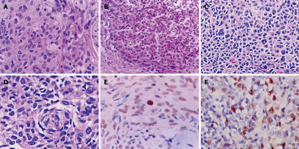
Figure 3 Atypical morphology of FDC sarcoma (A-D) and expression of p53 protein (E) and Epstein-Barr virus-encoded RNA (EBER) (F) in tumor cells.
A-D: Epithelioid (A and B) and pleomorphic tumor cells (C and D) are arranged in a sheet-like or diffuse pattern. Lymphocyte infiltration is less prominent (A and B) or absent (C and D) in these areas. HE: A, C and D, × 400; B, × 200; E: Nuclear immunoreactivity for p53 protein in majority of tumor cells. S-P, × 400; F: In situ hybridization signal for EBER in tumor cells, × 400.
- Citation: Li L, Shi YH, Guo ZJ, Qiu T, Guo L, Yang HY, Zhang X, Zhao XM, Su Q. Clinicopathological features and prognosis assessment of extranodal follicular dendritic cell sarcoma. World J Gastroenterol 2010; 16(20): 2504-2519
- URL: https://www.wjgnet.com/1007-9327/full/v16/i20/2504.htm
- DOI: https://dx.doi.org/10.3748/wjg.v16.i20.2504


