Copyright
©2010 Baishideng.
World J Gastroenterol. May 14, 2010; 16(18): 2298-2301
Published online May 14, 2010. doi: 10.3748/wjg.v16.i18.2298
Published online May 14, 2010. doi: 10.3748/wjg.v16.i18.2298
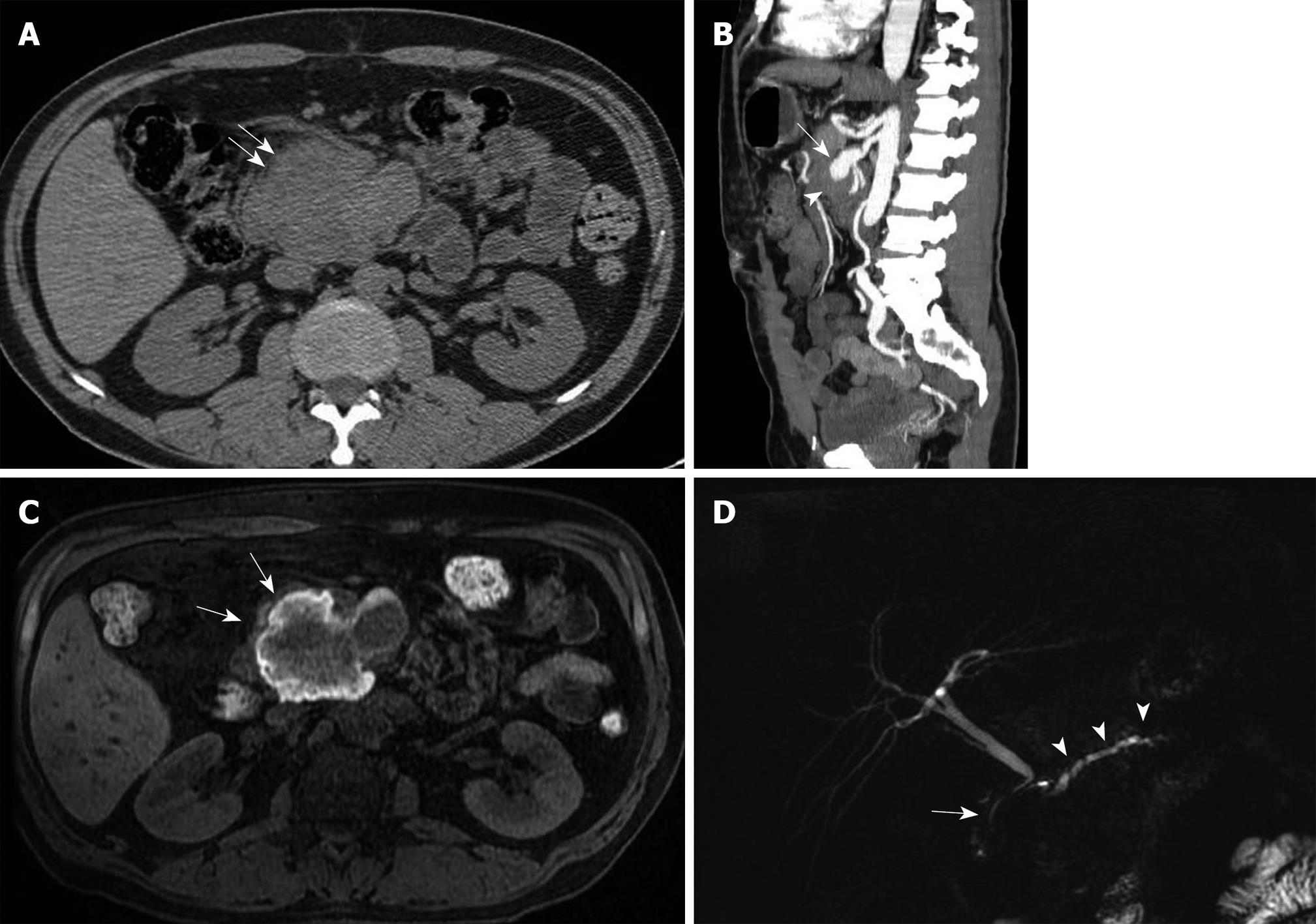
Figure 1 Images of the patient.
A: Axial unenhanced multi-detector computed tomography (MDCT) shows a soft-tissue mass (arrows) in the area of the uncinate process of the pancreas. There is no sign of calcification or fluid level; B: Sagittal multiplanar reconstruction clearly shows the superior mesenteric artery (arrow) involved in a giant soft-tissue attenuated mass - the hematoma (arrowhead) - at the pancreatic level. After contrast administration, there was no significant enhancement in the hematoma, due to its thrombotic content; C: Magnetic resonance, axial unenhanced gradient echo T1-weighted imaging with fat suppression. The T1 hyperintense perilesional signal (arrows) was caused by the blood content; D: Magnetic resonance cholangiopancreatography (MRCP) shows dilatation of the main pancreatic duct (arrowheads); the choledochus (arrow) was compressed and dislocated, with mild narrowing of the lumen in the distal tract.
- Citation: Palmucci S, Mauro LA, Milone P, Di Stefano F, Scolaro A, Di Cataldo A, Ettorre GC. Diagnosis of ruptured superior mesenteric artery aneurysm mimicking a pancreatic mass. World J Gastroenterol 2010; 16(18): 2298-2301
- URL: https://www.wjgnet.com/1007-9327/full/v16/i18/2298.htm
- DOI: https://dx.doi.org/10.3748/wjg.v16.i18.2298


