Copyright
©2010 Baishideng.
World J Gastroenterol. May 7, 2010; 16(17): 2109-2119
Published online May 7, 2010. doi: 10.3748/wjg.v16.i17.2109
Published online May 7, 2010. doi: 10.3748/wjg.v16.i17.2109
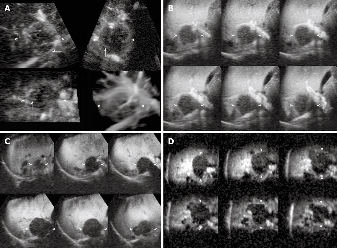
Figure 2 CE 3D US images of the liver in a 64-year-old woman with metastases from pancreatic carcinoma in the anterior superior segment of the right lobe.
A: Images in the early phase show peripheral ring-like enhancement with peritumoral vessels (arrows) in plane A (upper left), plane B (upper right), plane C (lower left) and the sonographic angiogram rendered by average intensity with surface mode (lower right); B: Tomographic ultrasound image with slice distance 2.0 mm in plane A shows peripheral ring-like enhancement in the middle phase; C, D: Tomographic ultrasound images (TUI) in plane A with slice distance 2.5 mm (C) and that in plane C with slice distance 2.0 mm (D) show hypoechoic pattern in the late phase. Enhancement change of washout was detected in this lesion. Arrowheads indicate the tumor margin.
- Citation: Luo W, Numata K, Morimoto M, Nozaki A, Ueda M, Kondo M, Morita S, Tanaka K. Differentiation of focal liver lesions using three-dimensional ultrasonography: Retrospective and prospective studies. World J Gastroenterol 2010; 16(17): 2109-2119
- URL: https://www.wjgnet.com/1007-9327/full/v16/i17/2109.htm
- DOI: https://dx.doi.org/10.3748/wjg.v16.i17.2109


