Copyright
©2010 Baishideng.
World J Gastroenterol. Apr 14, 2010; 16(14): 1720-1726
Published online Apr 14, 2010. doi: 10.3748/wjg.v16.i14.1720
Published online Apr 14, 2010. doi: 10.3748/wjg.v16.i14.1720
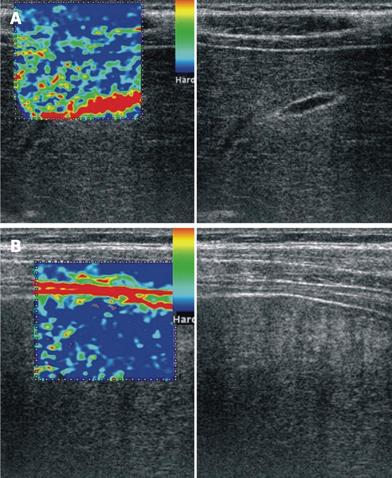
Figure 4 Real-time elastography images of right liver lobe.
A: 45-year-old patient with alcoholic liver steatosis. Region of interest include a large vessel, therefore, the liver parenchyma had a hard appearance (blue/green) in contrast with the extremely compressed (red) vessel; B: 52-year-old patient with liver cirrhosis and small amount of ascites surrounding the liver. There was very compressive and elastic fluid (red), and the liver parenchyma was depicted as very hard (blue). Consequently, other types of ascites might possibly induce similar artifacts, even in the presence of normal liver tissue.
- Citation: Gheonea DI, Săftoiu A, Ciurea T, Gorunescu F, Iordache S, Popescu GL, Belciug S, Gorunescu M, Săndulescu L. Real-time sono-elastography in the diagnosis of diffuse liver diseases. World J Gastroenterol 2010; 16(14): 1720-1726
- URL: https://www.wjgnet.com/1007-9327/full/v16/i14/1720.htm
- DOI: https://dx.doi.org/10.3748/wjg.v16.i14.1720


