Copyright
©2010 Baishideng.
World J Gastroenterol. Mar 14, 2010; 16(10): 1201-1208
Published online Mar 14, 2010. doi: 10.3748/wjg.v16.i10.1201
Published online Mar 14, 2010. doi: 10.3748/wjg.v16.i10.1201
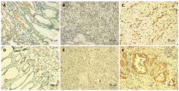
Figure 5 GATA-4 (A-C) or GATA-5 (D-F) protein in various gastric mucosa samples by immunohistochemical staining (IHC).
A and D: GATA-4 and GATA-5 expression were confined to the gland region in the normal gastric mucosa; B and E: Carcinoma cells containing methylated alleles of GATA-4 (G89) or GATA-5 (G48) exhibited negative staining; C and F: Unmethylated cancer cells exhibited positive staining in case G27 and G45, respectively. Diaminobenzidine was used as a chromogen, followed by counterstaining with hematoxylin. Black bar, 50 μm in length.
-
Citation: Wen XZ, Akiyama Y, Pan KF, Liu ZJ, Lu ZM, Zhou J, Gu LK, Dong CX, Zhu BD, Ji JF, You WC, Deng DJ. Methylation of
GATA-4 andGATA-5 and development of sporadic gastric carcinomas. World J Gastroenterol 2010; 16(10): 1201-1208 - URL: https://www.wjgnet.com/1007-9327/full/v16/i10/1201.htm
- DOI: https://dx.doi.org/10.3748/wjg.v16.i10.1201


