Copyright
©2010 Baishideng.
World J Gastroenterol. Jan 7, 2010; 16(1): 21-29
Published online Jan 7, 2010. doi: 10.3748/wjg.v16.i1.21
Published online Jan 7, 2010. doi: 10.3748/wjg.v16.i1.21
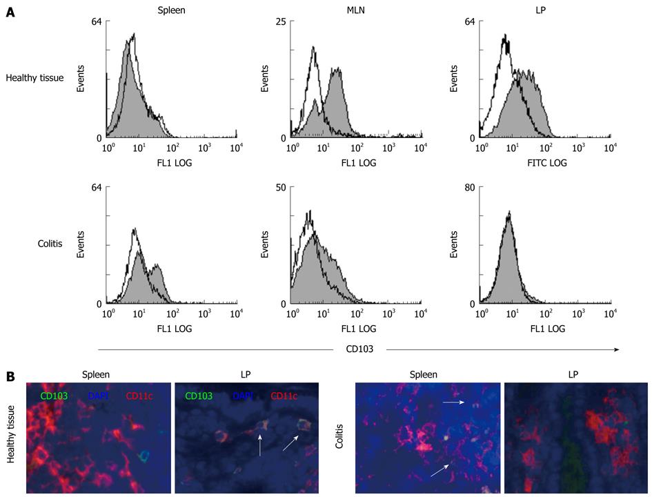
Figure 4 CD103+ DC are found in mucosal tissues of healthy mice and are lost during colitis.
A: Primary DC were isolated from different tissues (spleen, MLN, colonic LP) of healthy animals and mice with colitis. FACS analysis was performed after staining of CD11c+ DC with FITC-conjugated mAb against CD103, the integrin αEβ7. Data presented are representative of three independent experiments that were carried out with cells derived from animals with transfer colitis. Similar results were generated in the DSS colitis model; B: Tissue was harvested from spleen and LP of healthy animals and mice with colitis. Immunofluorescence was performed using an anti-CD11c+ mAb to stain for intestinal DC and an anti-CD103 mAb to recognize the integrin αEβ7. DAPI was used to visualize nuclei. Images show overlays of CD103 (green), CD11c (red) and DAPI (blue). DC coexpressing CD103 appear yellow (indicated by arrows). Representative sections from 5 mice per group are shown (magnification 100 ×). Staining was performed on tissue derived from colitic animals with transfer colitis as well as chronic DSS colitis and revealed similar results. Sections shown within this figure are derived from animals with transfer colitis.
- Citation: Strauch UG, Grunwald N, Obermeier F, Gürster S, Rath HC. Loss of CD103+ intestinal dendritic cells during colonic inflammation. World J Gastroenterol 2010; 16(1): 21-29
- URL: https://www.wjgnet.com/1007-9327/full/v16/i1/21.htm
- DOI: https://dx.doi.org/10.3748/wjg.v16.i1.21


