Copyright
©2010 Baishideng.
World J Gastroenterol. Jan 7, 2010; 16(1): 21-29
Published online Jan 7, 2010. doi: 10.3748/wjg.v16.i1.21
Published online Jan 7, 2010. doi: 10.3748/wjg.v16.i1.21
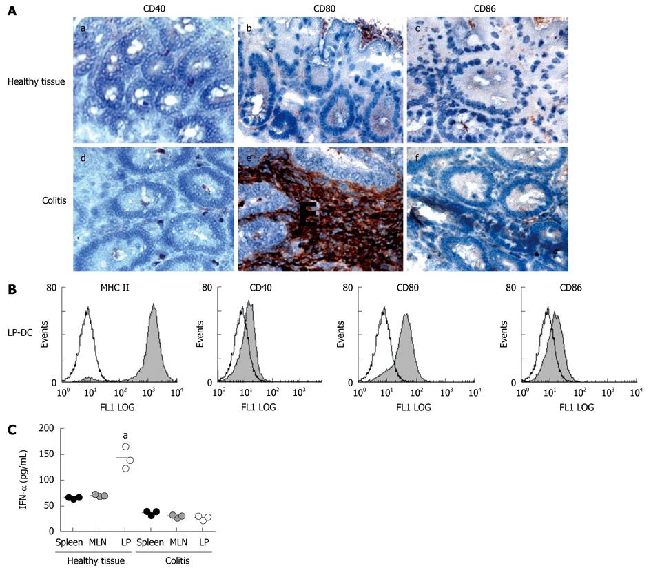
Figure 3 Intestinal DC are activated after induction of colitis.
A: Tissue was harvested from LP of healthy animals and from mice with colitis. Immunohistochemistry was performed with antibodies against the following costimulatory molecules: CD40 (a/d), CD80 (b/e), CD86 (c/f). Representative sections are shown (magnification 100 ×). Experiments were performed from 5 animals each group. Sections shown within this figure are derived from animals with transfer colitis, however staining was also performed on tissues derived from colitic animals with chronic DSS colitis and revealed similar results; B: LP-DC were isolated from inflamed intestinal tissue and FACS analysis was performed. CD11c+ DC were stained with FITC-conjugated mAbs for expression of MHC-class II and the costimulatory molecules CD40, CD80 and CD86. Data presented are representative of three independent experiments performed with cells derived from animals with transfer colitis. Similar results were generated in the DSS colitis model; C: Primary DC were isolated from different tissues (spleen, MLN, colonic LP) of healthy animals and mice with colitis. Isolated DC were incubated for 24 h in complete medium and levels of IFN-α were measured within the supernatants by ELISA (5 wells per situation). aP < 0.05. Data presented are from one of three independent experiments performed with cells derived from animals with transfer colitis. Similar results were generated in the DSS colitis model.
- Citation: Strauch UG, Grunwald N, Obermeier F, Gürster S, Rath HC. Loss of CD103+ intestinal dendritic cells during colonic inflammation. World J Gastroenterol 2010; 16(1): 21-29
- URL: https://www.wjgnet.com/1007-9327/full/v16/i1/21.htm
- DOI: https://dx.doi.org/10.3748/wjg.v16.i1.21


