Copyright
©2010 Baishideng.
World J Gastroenterol. Jan 7, 2010; 16(1): 131-135
Published online Jan 7, 2010. doi: 10.3748/wjg.v16.i1.131
Published online Jan 7, 2010. doi: 10.3748/wjg.v16.i1.131
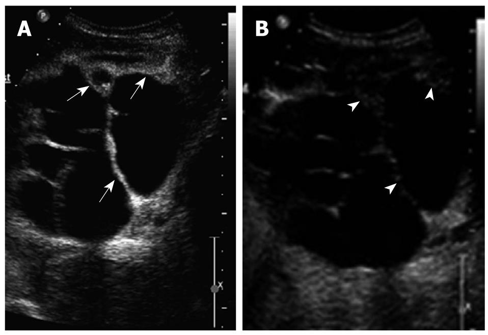
Figure 3 Biliary cystadenocarcinoma.
A: Contrast enhanced ultrasound (CEUS) shows hyperenhancement of the cystic wall, internal septations and intracystic solid portions during the arterial phase (arrows); B: CEUS shows hypoenhancement of the cystic wass, internal septations and intracystic solid portions during the portal and late phases (arrowheads).
- Citation: Ren XL, Yan RL, Yu XH, Zheng Y, Liu JE, Hou XB, Zuo SY, Fu XY, Chang H, Lu JH. Biliary cystadenocarcinoma diagnosed with real-time contrast-enhanced ultrasonography: Report of a case with diagnostic features. World J Gastroenterol 2010; 16(1): 131-135
- URL: https://www.wjgnet.com/1007-9327/full/v16/i1/131.htm
- DOI: https://dx.doi.org/10.3748/wjg.v16.i1.131


