Copyright
©2010 Baishideng.
World J Gastroenterol. Jan 7, 2010; 16(1): 126-130
Published online Jan 7, 2010. doi: 10.3748/wjg.v16.i1.126
Published online Jan 7, 2010. doi: 10.3748/wjg.v16.i1.126
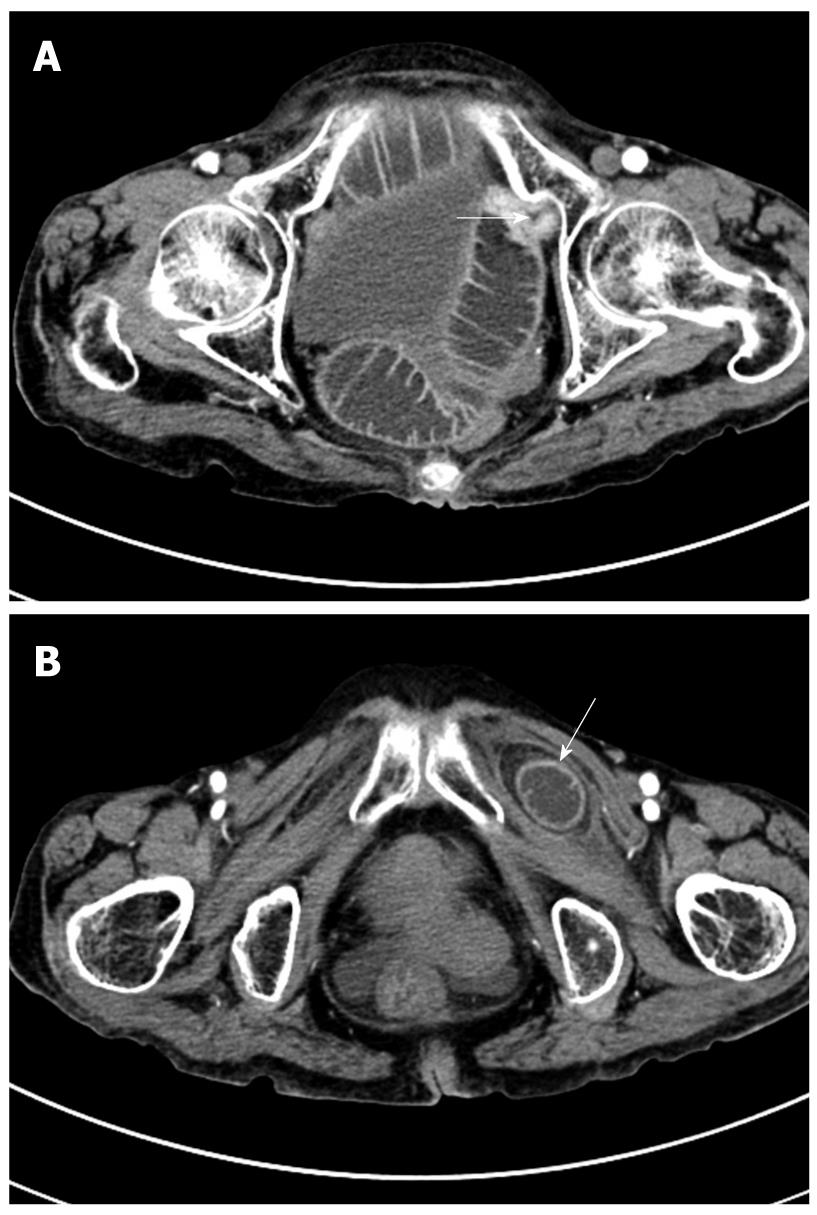
Figure 3 Abdominal axial CT image.
A: Severe dilated small bowel and abrupt stenosis at the terminal ileum in the pelvic cavity (arrow); B: A low-density mass with clear border located between the pectineus and the left external obturator muscles (arrow).
- Citation: Zhang H, Cong JC, Chen CS. Ileum perforation due to delayed operation in obturator hernia: A case report and review of literatures. World J Gastroenterol 2010; 16(1): 126-130
- URL: https://www.wjgnet.com/1007-9327/full/v16/i1/126.htm
- DOI: https://dx.doi.org/10.3748/wjg.v16.i1.126


