Copyright
©2009 The WJG Press and Baishideng.
World J Gastroenterol. Mar 7, 2009; 15(9): 1065-1071
Published online Mar 7, 2009. doi: 10.3748/wjg.15.1065
Published online Mar 7, 2009. doi: 10.3748/wjg.15.1065
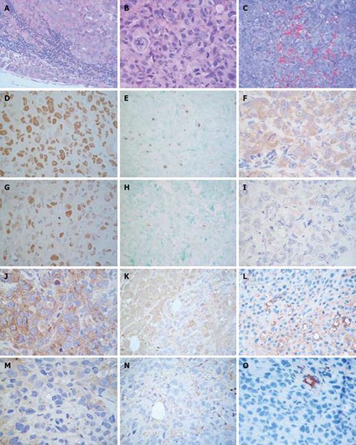
Figure 3 Histopathology and immunohistochemistry of PANC-1 xenografted mice.
Formalin fixed paraffin embedded tissue sections of control mice were stained with HE. A: Metastatic lymph node (× 400); B: Xenografts (× 630). Immunohistochemistry in control and AG treated mice. Tissue sections were stained with DAB and counterstained with hematoxylin; C: Formalin fixed paraffin embedded tissue sections of control mice were stained with trichromic solution (× 400); D, G: Ki-67 (× 630); E, H: Tunel (× 400); F, I: Bax (× 630); J, M: VEGF (× 630); K, N: eNOS (× 630); L, O: CD34 in control (× 400) and AG treated (× 630) mice. Formalin fixed paraffin embedded tissue sections of control mice were stained with trichrome solution.
- Citation: Mohamad NA, Cricco GP, Sambuco LA, Croci M, Medina VA, Gutiérrez AS, Bergoc RM, Rivera ES, Martín GA. Aminoguanidine impedes human pancreatic tumor growth and metastasis development in nude mice. World J Gastroenterol 2009; 15(9): 1065-1071
- URL: https://www.wjgnet.com/1007-9327/full/v15/i9/1065.htm
- DOI: https://dx.doi.org/10.3748/wjg.15.1065


