Copyright
©2009 The WJG Press and Baishideng.
World J Gastroenterol. Feb 14, 2009; 15(6): 675-683
Published online Feb 14, 2009. doi: 10.3748/wjg.15.675
Published online Feb 14, 2009. doi: 10.3748/wjg.15.675
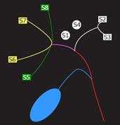
Figure 4 Normal anatomy of biliary drainage: Right posterior duct (RPD) and right anterior duct (RAD) drain, respectively, S6-S7 and S8-S5; right hepatic duct (RHD) is formed by confluence of RPD and RAD.
Left hepatic duct (LHD) drains S2-S3. S1-S4 can be drained by LHD or by RHD. The common hepatic duct (CHD) arises from the confluence of RHD and LHD.
- Citation: Caruso S, Miraglia R, Maruzzelli L, Gruttadauria S, Luca A, Gridelli B. Imaging in liver transplantation. World J Gastroenterol 2009; 15(6): 675-683
- URL: https://www.wjgnet.com/1007-9327/full/v15/i6/675.htm
- DOI: https://dx.doi.org/10.3748/wjg.15.675


