Copyright
©2009 The WJG Press and Baishideng.
World J Gastroenterol. Feb 7, 2009; 15(5): 615-621
Published online Feb 7, 2009. doi: 10.3748/wjg.15.615
Published online Feb 7, 2009. doi: 10.3748/wjg.15.615
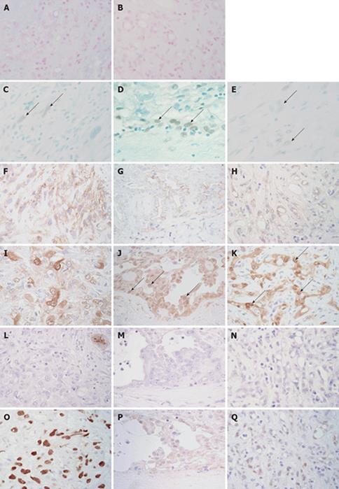
Figure 4 Immunohistochemical and special stains.
Alcian blue (pH 2.5) showed positive staining on the lumen of adenomatoid tumors (A). This positive staining was sensitive to digestion with hyaluronidase (B). Nuclear WT-1 immunoreactivity (arrows) was detected focally in sarcomatoid (C), papillary epithelioid (D) and microcystic (E) components. Immunostaining for WT-1 and methyl green. Sarcomatoid tumor cells showed focal membranous immunoreactivity for D2-40 (F). Membranous D2-40 immunoreactivity was detected in the papillary epithelioid (G) and microcystic (H) components. Immunostaining for D2-40 and hematoxylin. Sarcomatoid tumor cells showed focal and rather weak immunoreactivity for calretinin (I). Nuclear calretinin immunoreactivity (arrows) was detected in the papillary epithelioid (J) and microcystic (K) components. Immunostaining for calretinin and hematoxylin. Immunoreactivity for CA19-9 was not detected in sarcomatoid (L), papillary epithelioid (M) and microcystic (N) components. The apical surface of the entrapped bile duct showed immunoreactivity for CA19-9 (L, upper right corner). Immunostaining for CA19-9 and hematoxylin. Sarcomatoid (O) and microcystic (P) components showed strong nuclear immunoreactivity for p53. A few papillary epithelioid cells showed nuclear immunoreactivity for p53 (Q). (Immunostaining for p53 and hematoxylin, × 400).
- Citation: Sasaki M, Araki I, Yasui T, Kinoshita M, Itatsu K, Nojima T, Nakanuma Y. Primary localized malignant biphasic mesothelioma of the liver in a patient with asbestosis. World J Gastroenterol 2009; 15(5): 615-621
- URL: https://www.wjgnet.com/1007-9327/full/v15/i5/615.htm
- DOI: https://dx.doi.org/10.3748/wjg.15.615


