Copyright
©2009 The WJG Press and Baishideng.
World J Gastroenterol. Feb 7, 2009; 15(5): 615-621
Published online Feb 7, 2009. doi: 10.3748/wjg.15.615
Published online Feb 7, 2009. doi: 10.3748/wjg.15.615
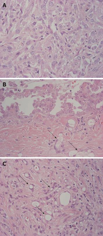
Figure 3 Histologic features of the tumor.
A: Sarcomatoid component was predominant and was composed of tumor cells with abundant eosinophilic or clear cytoplasm, indistinct cytoplasmic borders, round and atypical nuclei with vesicular fine chromatin and variably sized nucleoli; B: Papillary proliferation of epithelioid tumor cells was identified on the surface of the tumor. Epithelioid cells had eosinophilic cytoplasm with bland nuclei and distinct nucleoli. Arrows indicate microcystic (microglandular or adenomatoid) component; C: In the area near the surface, a microcystic (microglandular or adenomatoid) component showing microcystic structures with lace-like, adenoid cystic or signet ring appearance was detected (arrows) (HE × 400).
- Citation: Sasaki M, Araki I, Yasui T, Kinoshita M, Itatsu K, Nojima T, Nakanuma Y. Primary localized malignant biphasic mesothelioma of the liver in a patient with asbestosis. World J Gastroenterol 2009; 15(5): 615-621
- URL: https://www.wjgnet.com/1007-9327/full/v15/i5/615.htm
- DOI: https://dx.doi.org/10.3748/wjg.15.615


