Copyright
©2009 The WJG Press and Baishideng.
World J Gastroenterol. Feb 7, 2009; 15(5): 570-577
Published online Feb 7, 2009. doi: 10.3748/wjg.15.570
Published online Feb 7, 2009. doi: 10.3748/wjg.15.570
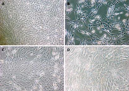
Figure 1 Morphological changes in human peritoneal mesothelial cells under phase contrast microscopy (× 40).
A: Morphology of mesothelial cells cultured in serum free DMEM; B: Exfoliation and naked areas of mesothelial cells after treatment with Astragalus injection; C: Morphological changes in mesothelial cells after treatment with 200 &mgr;g/mL Astragalus injection; D: No typical morphological changes in mesothelial cells after treatment with DMEM + 200 &mgr;g/mL Astragalus injection.
- Citation: Na D, Liu FN, Miao ZF, Du ZM, Xu HM. Astragalus extract inhibits destruction of gastric cancer cells to mesothelial cells by anti-apoptosis. World J Gastroenterol 2009; 15(5): 570-577
- URL: https://www.wjgnet.com/1007-9327/full/v15/i5/570.htm
- DOI: https://dx.doi.org/10.3748/wjg.15.570


