Copyright
©2009 The WJG Press and Baishideng.
World J Gastroenterol. Nov 21, 2009; 15(43): 5489-5492
Published online Nov 21, 2009. doi: 10.3748/wjg.15.5489
Published online Nov 21, 2009. doi: 10.3748/wjg.15.5489
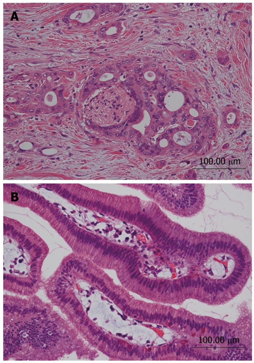
Figure 6 Histological appearance of two lesions (HE).
A: Intraductal papillary mucinous adenoma; B: Moderately differentiated tubular adenocarcinoma.
- Citation: Sakamoto H, Kitano M, Komaki T, Imai H, Kamata K, Kimura M, Takeyama Y, Kudo M. Small invasive ductal carcinoma of the pancreas distinct from branch duct intraductal papillary mucinous neoplasm. World J Gastroenterol 2009; 15(43): 5489-5492
- URL: https://www.wjgnet.com/1007-9327/full/v15/i43/5489.htm
- DOI: https://dx.doi.org/10.3748/wjg.15.5489


