Copyright
©2009 The WJG Press and Baishideng.
World J Gastroenterol. Nov 21, 2009; 15(43): 5489-5492
Published online Nov 21, 2009. doi: 10.3748/wjg.15.5489
Published online Nov 21, 2009. doi: 10.3748/wjg.15.5489
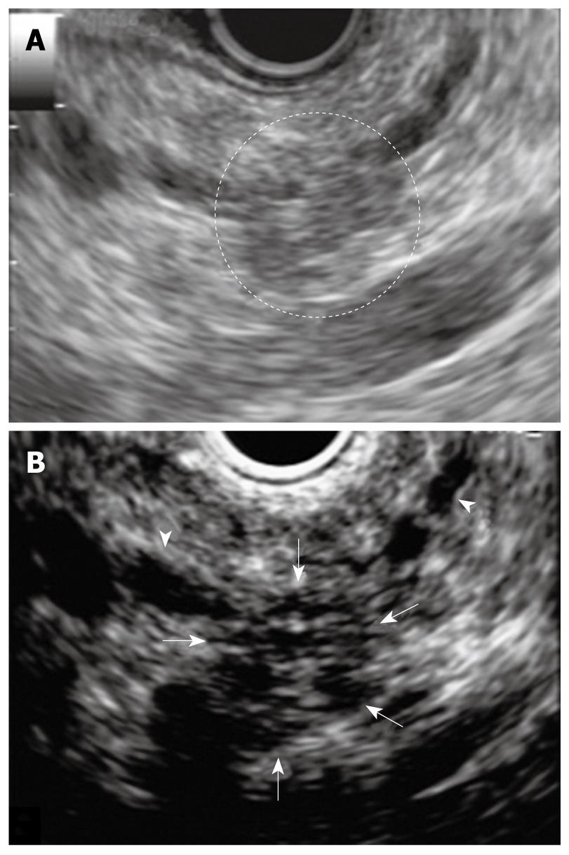
Figure 4 Endoscopic ultrasonography (EUS) at two years later examination showing pancreatic tail.
A: EUS showing low echoic lesion which is vague and the area had unclear margins in the pancreatic tail; B: Contrast-enhanced harmonic EUS showing a clear margin of 10 mm in diameter and hypovascular nodule compared with surrounding pancreatic tissue (arrows). MPD: Main pancreatic duct (arrowheads).
- Citation: Sakamoto H, Kitano M, Komaki T, Imai H, Kamata K, Kimura M, Takeyama Y, Kudo M. Small invasive ductal carcinoma of the pancreas distinct from branch duct intraductal papillary mucinous neoplasm. World J Gastroenterol 2009; 15(43): 5489-5492
- URL: https://www.wjgnet.com/1007-9327/full/v15/i43/5489.htm
- DOI: https://dx.doi.org/10.3748/wjg.15.5489


