Copyright
©2009 The WJG Press and Baishideng.
World J Gastroenterol. Nov 21, 2009; 15(43): 5418-5424
Published online Nov 21, 2009. doi: 10.3748/wjg.15.5418
Published online Nov 21, 2009. doi: 10.3748/wjg.15.5418
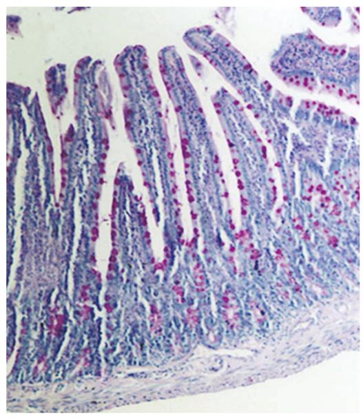
Figure 1 Photomicrograph of the tunica mucosa (M) and muscularis mucosa (MM) of the small intestine in the GH-administered group.
Villous hypertrophy and increased goblet cells were seen in the small intestine. PAS, × 100 (Original magnification).
- Citation: Ersoy B, Ozbilgin K, Kasirga E, Inan S, Coskun S, Tuglu I. Effect of growth hormone on small intestinal homeostasis relation to cellular mediators IGF-I and IGFBP-3. World J Gastroenterol 2009; 15(43): 5418-5424
- URL: https://www.wjgnet.com/1007-9327/full/v15/i43/5418.htm
- DOI: https://dx.doi.org/10.3748/wjg.15.5418


