Copyright
©2009 The WJG Press and Baishideng.
World J Gastroenterol. Nov 14, 2009; 15(42): 5360-5363
Published online Nov 14, 2009. doi: 10.3748/wjg.15.5360
Published online Nov 14, 2009. doi: 10.3748/wjg.15.5360
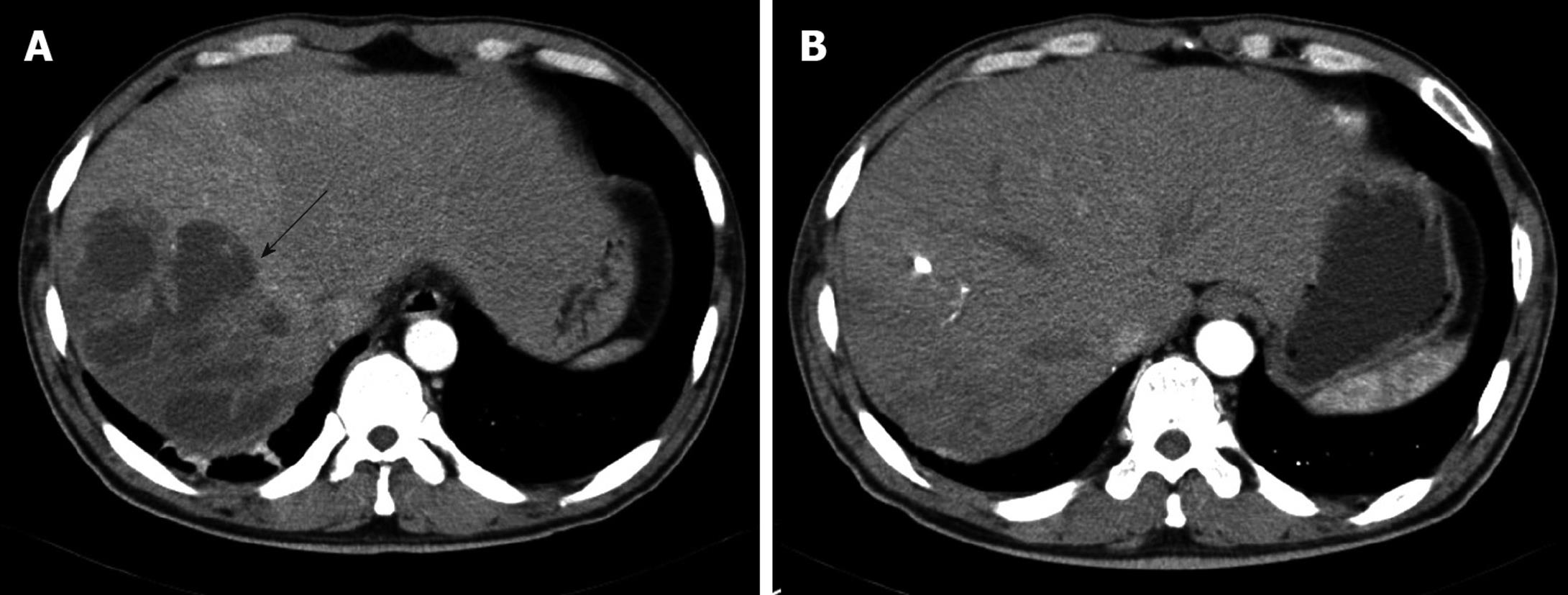
Figure 1 Contrast-enhanced computed tomography (CT) of the liver.
A: Abdominal CT scan showing a multi-loculated liver abscess (12 cm × 9 cm) in the posterior inferior (VI segment) and posterior superior segment of the right lobe (VII segment) of the liver (arrow); B: On hospital day 30, the liver abscess was almost absorbed.
-
Citation: Na JS, Kim TH, Kim HS, Park SH, Song HS, Cha SW, Yoon HJ. Liver abscess and sepsis with
Bacillus pantothenticus in an immunocompetent patient: A first case report. World J Gastroenterol 2009; 15(42): 5360-5363 - URL: https://www.wjgnet.com/1007-9327/full/v15/i42/5360.htm
- DOI: https://dx.doi.org/10.3748/wjg.15.5360


