Copyright
©2009 The WJG Press and Baishideng.
World J Gastroenterol. Nov 7, 2009; 15(41): 5232-5235
Published online Nov 7, 2009. doi: 10.3748/wjg.15.5232
Published online Nov 7, 2009. doi: 10.3748/wjg.15.5232
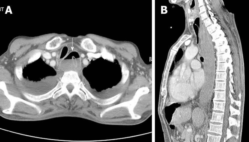
Figure 2 Chest CT images.
A: The transverse view of the proximal esophagus shows a concentric intramural hematoma and mucosal dissection with an air-fluid level in the false lumen (arrow). Bilateral pleural effusions are seen; B: The sagittal view shows an extensive intramural hematoma of the esophagus.
- Citation: Shim J, Jang JY, Hwangbo Y, Dong SH, Oh JH, Kim HJ, Kim BH, Chang YW, Chang R. Recurrent massive bleeding due to dissecting intramural hematoma of the esophagus: Treatment with therapeutic angiography. World J Gastroenterol 2009; 15(41): 5232-5235
- URL: https://www.wjgnet.com/1007-9327/full/v15/i41/5232.htm
- DOI: https://dx.doi.org/10.3748/wjg.15.5232


