Copyright
©2009 The WJG Press and Baishideng.
World J Gastroenterol. Nov 7, 2009; 15(41): 5165-5175
Published online Nov 7, 2009. doi: 10.3748/wjg.15.5165
Published online Nov 7, 2009. doi: 10.3748/wjg.15.5165
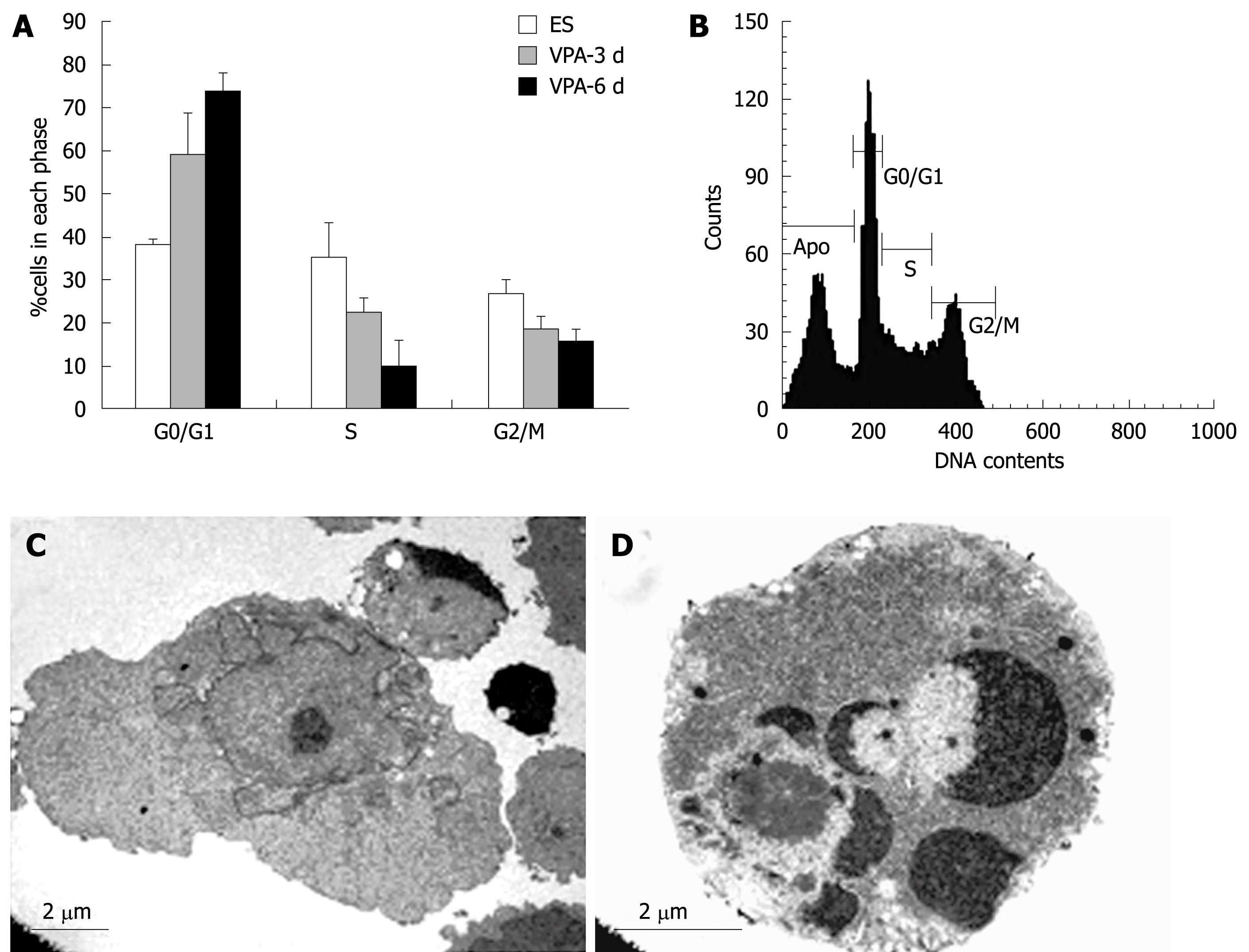
Figure 6 VPA-induced cell cycle arrest and apoptosis during hepatic differentiation.
A: Cell cycle analysis revealed that exposure to VPA decreased the proportion of cells in S phase and increased the proportion of cells in the G0/G1 phase. Approximately 75% of the cells were arrested in the G0/G1 phase and only 10% of the cells in the S phase after 6 d of treatment with 1 mmol/L VPA, whereas greater than 37% of control ES cells were in the S phase; B: The analysis of apoptosis proportions during 3 d of treatment with VPA; C, D: Ultrastructural observations showed that some cells presented typical apoptotic morphology when treated with VPA for more than 5 d.
- Citation: Dong XJ, Zhang GR, Zhou QJ, Pan RL, Chen Y, Xiang LX, Shao JZ. Direct hepatic differentiation of mouse embryonic stem cells induced by valproic acid and cytokines. World J Gastroenterol 2009; 15(41): 5165-5175
- URL: https://www.wjgnet.com/1007-9327/full/v15/i41/5165.htm
- DOI: https://dx.doi.org/10.3748/wjg.15.5165


