Copyright
©2009 The WJG Press and Baishideng.
World J Gastroenterol. Oct 21, 2009; 15(39): 4907-4914
Published online Oct 21, 2009. doi: 10.3748/wjg.15.4907
Published online Oct 21, 2009. doi: 10.3748/wjg.15.4907
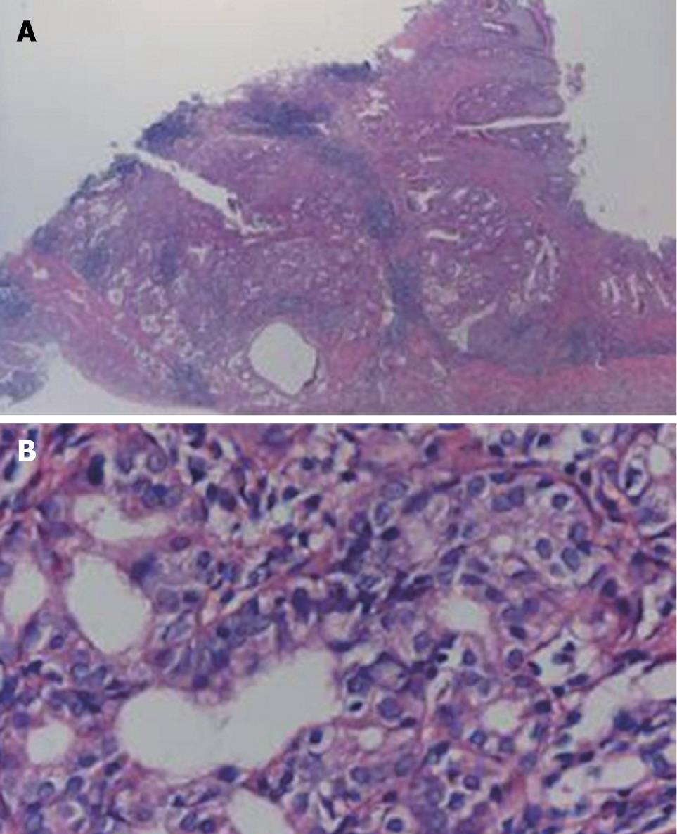
Figure 2 Typical adenocarcinoma in the pyloric mucosa of H pylori-infected MGs.
Shown is a typical well-differentiated adenocarcinoma (A, B) stained with HE. Images were obtained at × 100 (A) and × 400 (B).
- Citation: Kuo CH, Hu HM, Tsai PY, Wu IC, Yang SF, Chang LL, Wang JY, Jan CM, Wang WM, Wu DC. Short-term Celecoxib intervention is a safe and effective chemopreventive for gastric carcinogenesis based on a Mongolian gerbil model. World J Gastroenterol 2009; 15(39): 4907-4914
- URL: https://www.wjgnet.com/1007-9327/full/v15/i39/4907.htm
- DOI: https://dx.doi.org/10.3748/wjg.15.4907


