Copyright
©2009 The WJG Press and Baishideng.
World J Gastroenterol. Sep 28, 2009; 15(36): 4576-4581
Published online Sep 28, 2009. doi: 10.3748/wjg.15.4576
Published online Sep 28, 2009. doi: 10.3748/wjg.15.4576
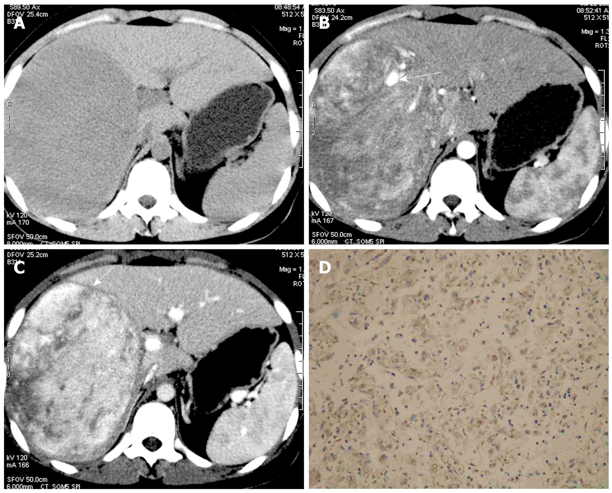
Figure 3 17-year-old girl with epithelioid angiomyolipoma in right lobe of liver (the patient had a history of tuberous sclerosis complex, patient 5).
A: Non-enhanced CT scan shows homogeneous hypoattenuating lesion in segment V/VII/VIII; B: Contrast-enhanced CT scan shows inhomogeneous enhanced lesion with opacification of central vessels (arrow) on the arterial phase; C: The slight enhanced outer rim (arrow) around the inhomogeneous enhancement lesions is shown on portal phase contrast-enhanced CT; D: Immunohistochemical staining for HMB-45 shows diffusely positive staining in tumor cells within the cytoplasm (EnVision, × 100).
- Citation: Xu PJ, Shan Y, Yan FH, Ji Y, Ding Y, Zhou ML. Epithelioid angiomyolipoma of the liver: Cross-sectional imaging findings of 10 immunohistochemically-verified cases. World J Gastroenterol 2009; 15(36): 4576-4581
- URL: https://www.wjgnet.com/1007-9327/full/v15/i36/4576.htm
- DOI: https://dx.doi.org/10.3748/wjg.15.4576


