
















©2009 The WJG Press and Baishideng.
World J Gastroenterol. Sep 21, 2009; 15(35): 4461-4463
Published online Sep 21, 2009. doi: 10.3748/wjg.15.4461
Published online Sep 21, 2009. doi: 10.3748/wjg.15.4461
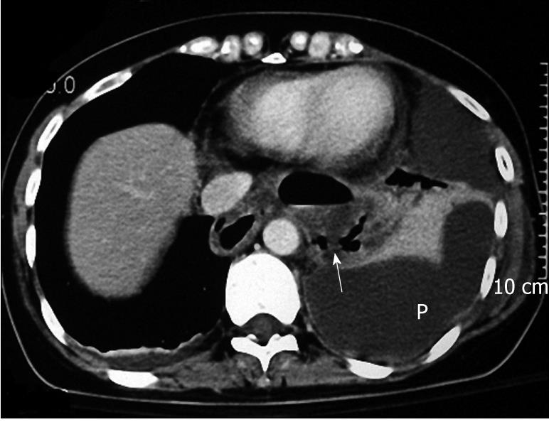
Figure 3 CT revealed esophageal dilatation and a rupture of the lower esophagus (arrow) with prominent pleural effusion (P) and fluid collection over the middle and lower mediastinum.
- Citation: Lin MT, Tai WC, Chiu KW, Chou YP, Tsai MC, Hu TH, Lee CM, Changchien CS, Chuah SK. Delayed presentation of intrathoracic esophageal perforation after pneumatic dilation for achalasia. World J Gastroenterol 2009; 15(35): 4461-4463
- URL: https://www.wjgnet.com/1007-9327/full/v15/i35/4461.htm
- DOI: https://dx.doi.org/10.3748/wjg.15.4461