Copyright
©2009 The WJG Press and Baishideng.
World J Gastroenterol. Aug 28, 2009; 15(32): 4049-4054
Published online Aug 28, 2009. doi: 10.3748/wjg.15.4049
Published online Aug 28, 2009. doi: 10.3748/wjg.15.4049
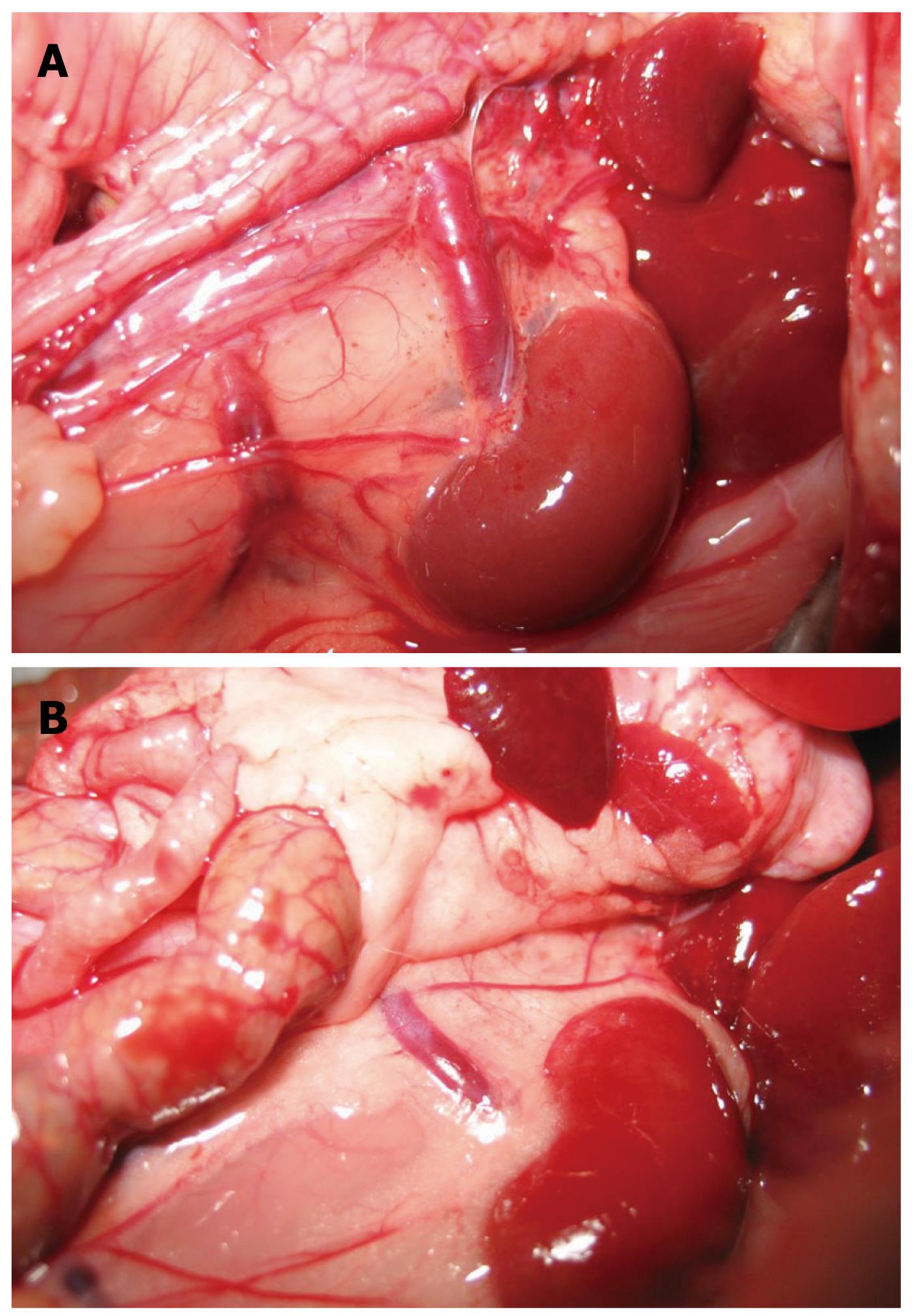
Figure 3 Collaterals after PVL.
A: In the control rat, the left renal vein was thin and of normal size, no collateral vessels could be seen between the spleen and the left kidney; B: Collaterals could be seen mainly between the spleen and the left kidney 10 wk after PVL. The left adrenal vein was markedly engorged. The collaterals could also be seen between the inferior mesenteric vein and the posterior peritoneum.
- Citation: Wen Z, Zhang JZ, Xia HM, Yang CX, Chen YJ. Stability of a rat model of prehepatic portal hypertension caused by partial ligation of the portal vein. World J Gastroenterol 2009; 15(32): 4049-4054
- URL: https://www.wjgnet.com/1007-9327/full/v15/i32/4049.htm
- DOI: https://dx.doi.org/10.3748/wjg.15.4049


