Copyright
©2009 The WJG Press and Baishideng.
World J Gastroenterol. Aug 28, 2009; 15(32): 4016-4022
Published online Aug 28, 2009. doi: 10.3748/wjg.15.4016
Published online Aug 28, 2009. doi: 10.3748/wjg.15.4016
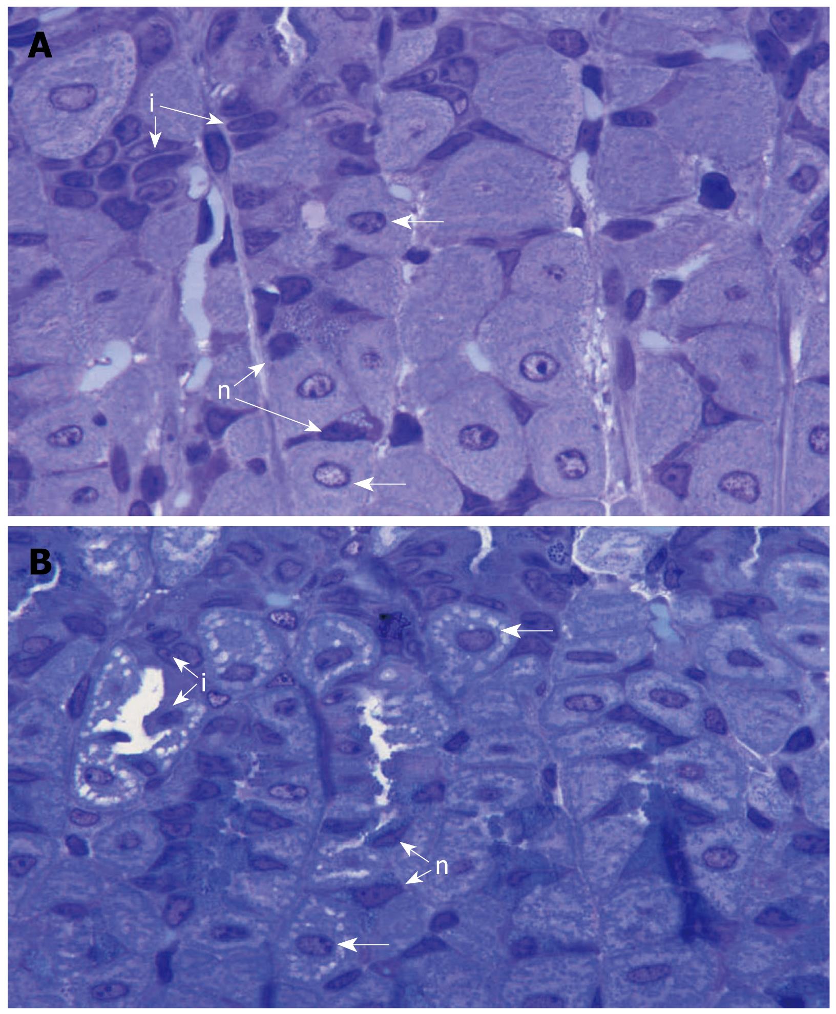
Figure 5 Semithin (0.
5-micron-thick) sections of the gastric mucosae of control (A) and smoke-treated (B) mice stained with toluidine blue to demonstrate the isthmus and neck regions of the gastric glands. The large numerous parietal cells (horizontal arrows) are separated by progenitor or isthmal cells (i) and neck cells (n). Note that, in the smoke-treated tissue but not control tissue, there are pale areas in the cytoplasm of parietal cells which represent expanded lumen of the intracellular canaliculi, × 1000.
- Citation: Hammadi M, Adi M, John R, Khoder GA, Karam SM. Dysregulation of gastric H,K-ATPase by cigarette smoke extract. World J Gastroenterol 2009; 15(32): 4016-4022
- URL: https://www.wjgnet.com/1007-9327/full/v15/i32/4016.htm
- DOI: https://dx.doi.org/10.3748/wjg.15.4016


