Copyright
©2009 The WJG Press and Baishideng.
World J Gastroenterol. Aug 28, 2009; 15(32): 3992-3998
Published online Aug 28, 2009. doi: 10.3748/wjg.15.3992
Published online Aug 28, 2009. doi: 10.3748/wjg.15.3992
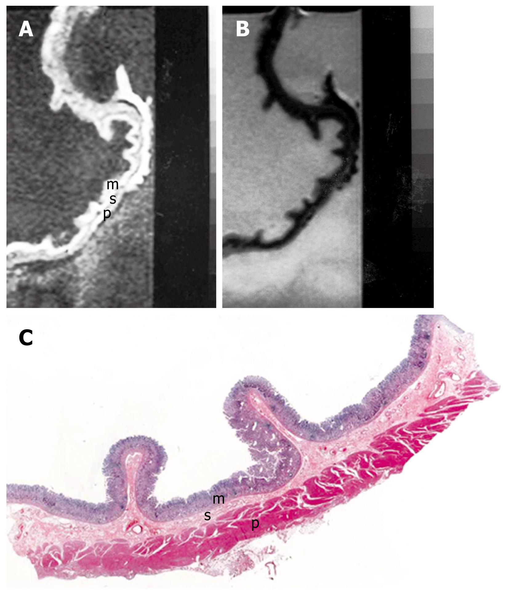
Figure 1 MRI and histology of normal gastric wall.
A: T1-weighted (500/20) sagittal image of resected gastric wall showed three layers. The inner layer corresponds to the mucosa (m) and the middle layer to the submucosa (s). The outer layer basically consists of the muscularis propria (p) from which the serosa cannot be differentiated; B: T2-weighted (2500/90) MRI showed low SI on mucosa and muscularis propria and relatively high SI on submucosa; C: Light microscopic section of normal gastric wall obtained from the greater curvature site of stomach body showed three layers which are compatible with inner mucosal layer (m), middle submucosa layer (s) and outer muscularis propria and serosal layer (p) (HE stain; original magnification, × 1).
- Citation: Kim IY, Kim SW, Shin HC, Lee MS, Jeong DJ, Kim CJ, Kim YT. MRI of gastric carcinoma: Results of T and N-staging in an in vitro study. World J Gastroenterol 2009; 15(32): 3992-3998
- URL: https://www.wjgnet.com/1007-9327/full/v15/i32/3992.htm
- DOI: https://dx.doi.org/10.3748/wjg.15.3992


