Copyright
©2009 The WJG Press and Baishideng.
World J Gastroenterol. Aug 21, 2009; 15(31): 3957-3959
Published online Aug 21, 2009. doi: 10.3748/wjg.15.3957
Published online Aug 21, 2009. doi: 10.3748/wjg.15.3957
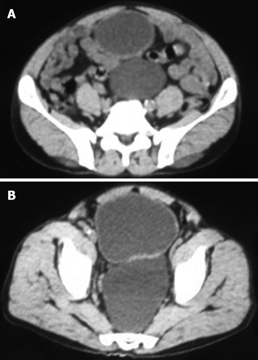
Figure 1 Computed tomography showed a 15 cm × 10 cm low density cystic lesion with smooth contours located in the presacral region, pushing the rectum to the right and the sigmoid colon and bladder superiorly (A and B).
- Citation: Akbulut S, Cakabay B, Sezgin A, Isen K, Senol A. Giant vesical diverticulum: A rare cause of defecation disturbance. World J Gastroenterol 2009; 15(31): 3957-3959
- URL: https://www.wjgnet.com/1007-9327/full/v15/i31/3957.htm
- DOI: https://dx.doi.org/10.3748/wjg.15.3957


