
















©2009 The WJG Press and Baishideng.
World J Gastroenterol. Jan 21, 2009; 15(3): 289-299
Published online Jan 21, 2009. doi: 10.3748/wjg.15.289
Published online Jan 21, 2009. doi: 10.3748/wjg.15.289
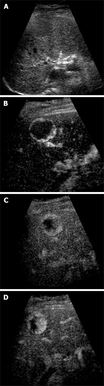
Figure 2 Haemangioma.
A: Baseline focal hypo-echoic lesion; B: Arterial phase showing peripheral rim enhancement; C: Portal venous phase showing peripheral globular rim enhancement; D: Late phase showing progressive centripetal filling-in which is characteristic for hemangioma.
- Citation: Leen E, Kumar S, Khan SA, Low G, Ong KO, Tait P, Averkiou M. Contrast-enhanced 3D ultrasound in the radiofrequency ablation of liver tumors. World J Gastroenterol 2009; 15(3): 289-299
- URL: https://www.wjgnet.com/1007-9327/full/v15/i3/289.htm
- DOI: https://dx.doi.org/10.3748/wjg.15.289