Copyright
©2009 The WJG Press and Baishideng.
World J Gastroenterol. Aug 7, 2009; 15(29): 3691-3693
Published online Aug 7, 2009. doi: 10.3748/wjg.15.3691
Published online Aug 7, 2009. doi: 10.3748/wjg.15.3691
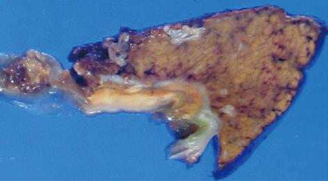
Figure 3 Cut surface of the specimen.
A yellowish tumor about 2 cm in diameter was observed between the mucosa of the body of the gallbladder and the liver parenchyma in the cut specimen.
- Citation: Makino I, Yamaguchi T, Sato N, Yasui T, Kita I. Xanthogranulomatous cholecystitis mimicking gallbladder carcinoma with a false-positive result on fluorodeoxyglucose PET. World J Gastroenterol 2009; 15(29): 3691-3693
- URL: https://www.wjgnet.com/1007-9327/full/v15/i29/3691.htm
- DOI: https://dx.doi.org/10.3748/wjg.15.3691


