Copyright
©2009 The WJG Press and Baishideng.
World J Gastroenterol. Aug 7, 2009; 15(29): 3664-3669
Published online Aug 7, 2009. doi: 10.3748/wjg.15.3664
Published online Aug 7, 2009. doi: 10.3748/wjg.15.3664
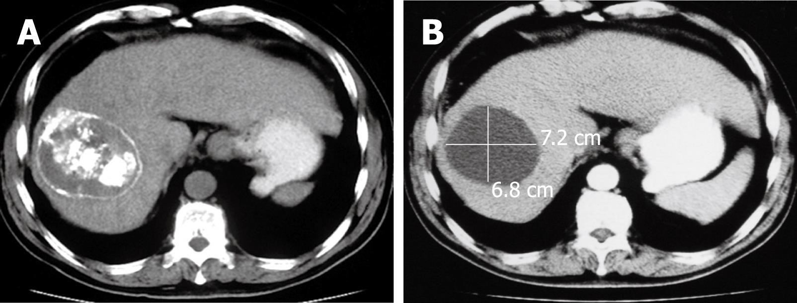
Figure 3 Histology showing the HCC mass at 3 mo (A) and 28 mo (B) after sequential therapy.
The cryoablated mass had cavitation formation. No cancer cells were identified by needle aspiration.
- Citation: Xu KC, Niu LZ, Zhou Q, Hu YZ, Guo DH, Liu ZP, Lan B, Mu F, Li YF, Zuo JS. Sequential use of transarterial chemoembolization and percutaneous cryosurgery for hepatocellular carcinoma. World J Gastroenterol 2009; 15(29): 3664-3669
- URL: https://www.wjgnet.com/1007-9327/full/v15/i29/3664.htm
- DOI: https://dx.doi.org/10.3748/wjg.15.3664


