Copyright
©2009 The WJG Press and Baishideng.
World J Gastroenterol. Jul 14, 2009; 15(26): 3303-3308
Published online Jul 14, 2009. doi: 10.3748/wjg.15.3303
Published online Jul 14, 2009. doi: 10.3748/wjg.15.3303
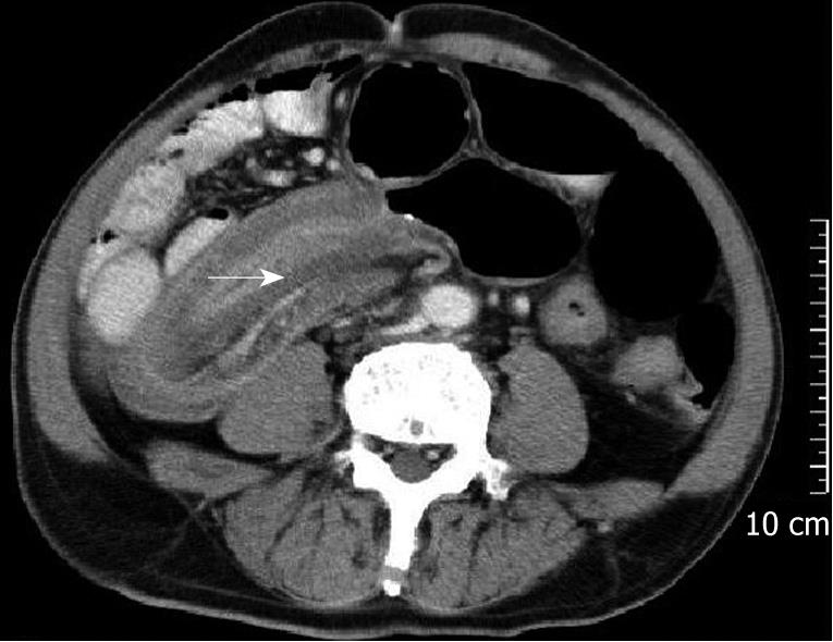
Figure 2 A 64-year-old man with an ileocolic intussusception due to a ileum B cell malignant lymphoma.
A sausage-shaped mass with high density soft tissue above represents the edematous bowel wall of the intussuscipiens and the intussusceptum, with fat density below, representing mesenteric fat. The higher linear density within the mesenteric fat (arrow) is mesenteric blood vessels. This appearance is caused by the axis of the intussusception being parallel with the computed tomography (CT) beam.
- Citation: Wang N, Cui XY, Liu Y, Long J, Xu YH, Guo RX, Guo KJ. Adult intussusception: A retrospective review of 41 cases. World J Gastroenterol 2009; 15(26): 3303-3308
- URL: https://www.wjgnet.com/1007-9327/full/v15/i26/3303.htm
- DOI: https://dx.doi.org/10.3748/wjg.15.3303


