Copyright
©2009 The WJG Press and Baishideng.
World J Gastroenterol. Jun 28, 2009; 15(24): 3065-3067
Published online Jun 28, 2009. doi: 10.3748/wjg.15.3065
Published online Jun 28, 2009. doi: 10.3748/wjg.15.3065
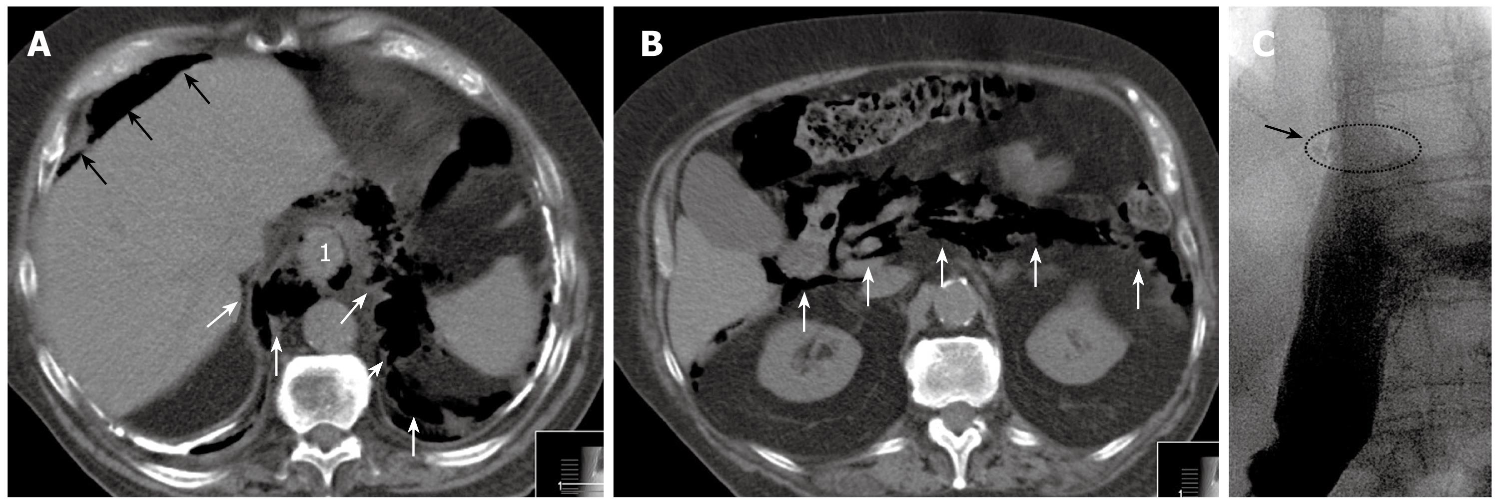
Figure 1 Pre- and postoperative radiographs.
A and B: CT scan of the abdomen revealing free intra-abdominal air (A, black arrows) and massive mediastinal (A, white arrows) and retroperitoneal (B, white arrows) air. 1Depicts the tumor localization at the GE junction; C: Water-soluble contrast medium swallowed after GE reconstruction (end-to-side esophago-gastrostomy) without evidence of anastomotic stenosis or leakage. The circle (arrow) points to the level of the anastomosis.
- Citation: Gillen S, Friess H, Kleeff J. Palliative cardia resection with gastroesophageal reconstruction for perforated carcinoma of the gastroesophageal junction. World J Gastroenterol 2009; 15(24): 3065-3067
- URL: https://www.wjgnet.com/1007-9327/full/v15/i24/3065.htm
- DOI: https://dx.doi.org/10.3748/wjg.15.3065


