Copyright
©2009 The WJG Press and Baishideng.
World J Gastroenterol. Jun 28, 2009; 15(24): 3003-3008
Published online Jun 28, 2009. doi: 10.3748/wjg.15.3003
Published online Jun 28, 2009. doi: 10.3748/wjg.15.3003
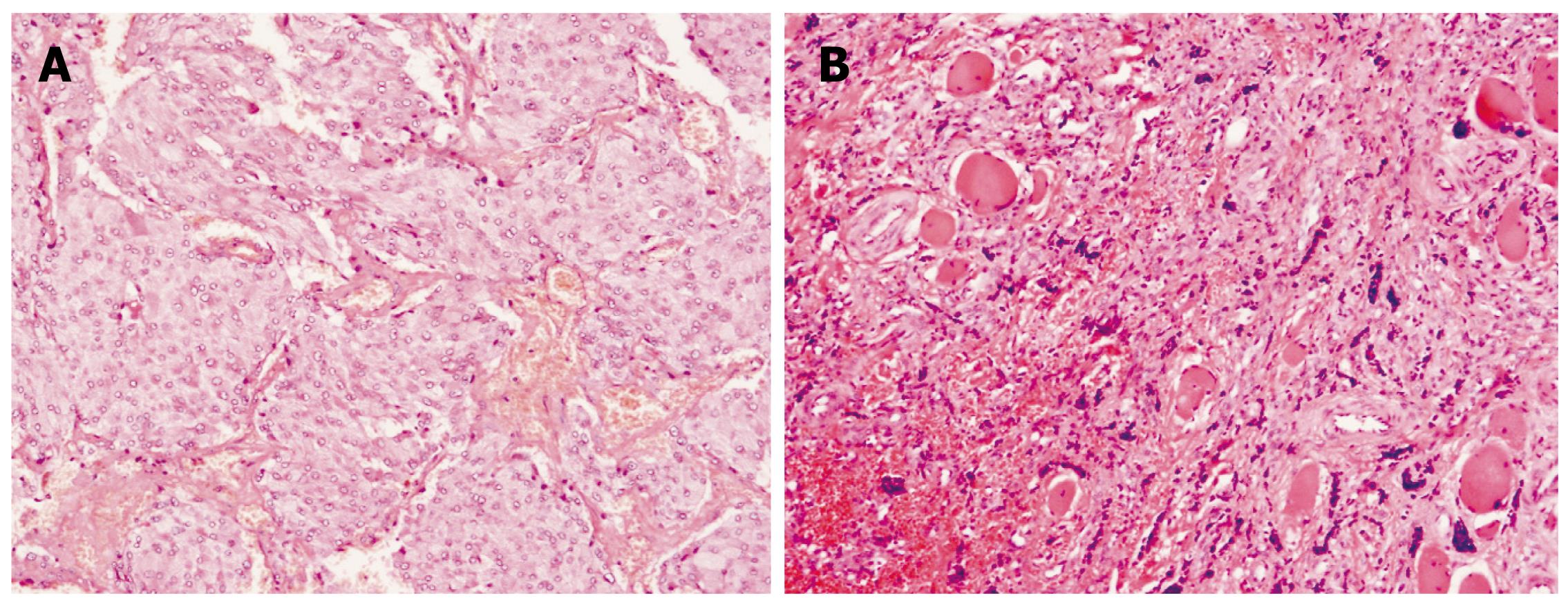
Figure 2 The histologic analysis of paraganglioma.
The hematoxylin-eosin stain of chief (type 1) cells arranged in one of the typical cell nests in a retroperitoneal benign tumor (A), and in a femur metastasis (B), (× 50).
- Citation: Feng N, Zhang WY, Wu XT. Clinicopathological analysis of paraganglioma with literature review. World J Gastroenterol 2009; 15(24): 3003-3008
- URL: https://www.wjgnet.com/1007-9327/full/v15/i24/3003.htm
- DOI: https://dx.doi.org/10.3748/wjg.15.3003


