Copyright
©2009 The WJG Press and Baishideng.
World J Gastroenterol. Jun 7, 2009; 15(21): 2657-2664
Published online Jun 7, 2009. doi: 10.3748/wjg.15.2657
Published online Jun 7, 2009. doi: 10.3748/wjg.15.2657
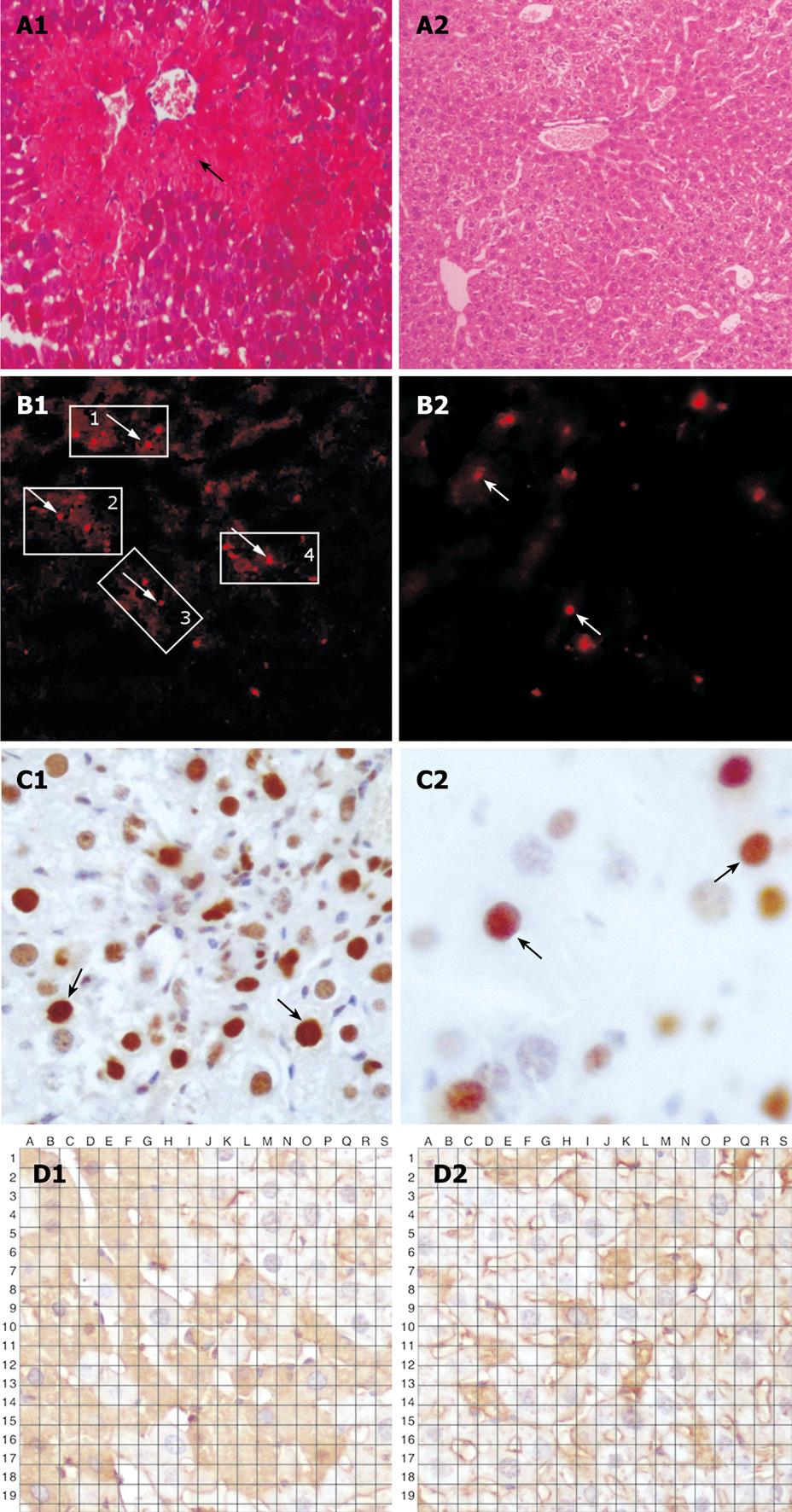
Figure 1 Histopathology of hepatic tissue from the two groups.
A: PKH26-labeled cells detected after establishment of acute liver failure animal model with extensive vacuolar degeneration and edema of hepatocytes in acute liver failure (A1) and normal liver tissue (A2); B: Sporadic PKH26-labeled bone marrow stem cells in experimental group (B1) and control group (B2); C: Expression of PCNA in sporadic PKH26-labeled bone marrow stem cells in experimental group (C1) and control group (C2); D: Expression of albumin and sporadic PKH26-labeled bone marrow stem cells in experimental group (D1) and control group (D2) ( × 200).
- Citation: Jin SZ, Meng XW, Han MZ, Sun X, Sun LY, Liu BR. Stromal cell derived factor-1 enhances bone marrow mononuclear cell migration in mice with acute liver failure. World J Gastroenterol 2009; 15(21): 2657-2664
- URL: https://www.wjgnet.com/1007-9327/full/v15/i21/2657.htm
- DOI: https://dx.doi.org/10.3748/wjg.15.2657


