Copyright
©2009 The WJG Press and Baishideng.
World J Gastroenterol. May 28, 2009; 15(20): 2433-2440
Published online May 28, 2009. doi: 10.3748/wjg.15.2433
Published online May 28, 2009. doi: 10.3748/wjg.15.2433
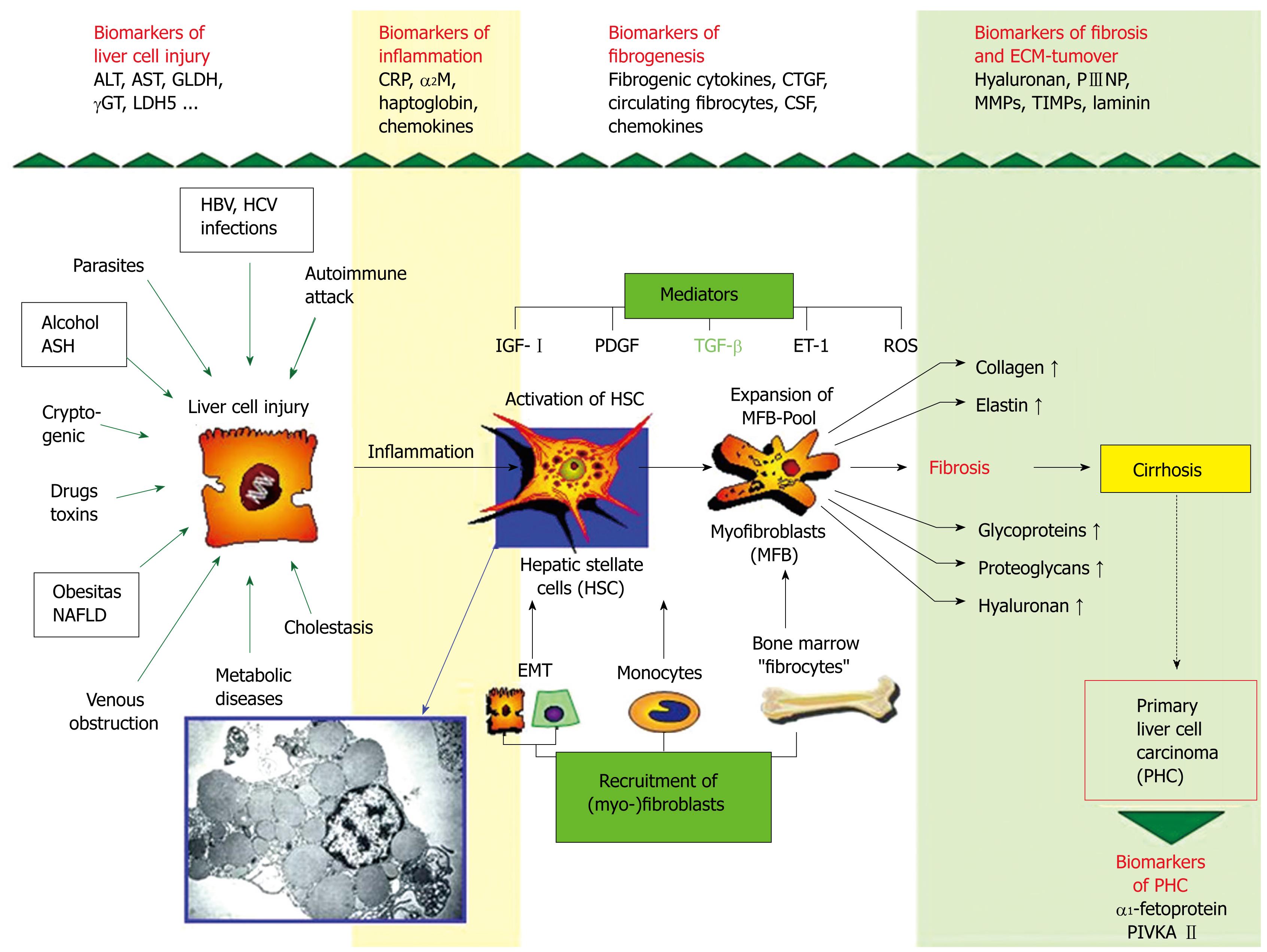
Figure 2 Synopsis of pathogenetic mechanisms of liver fibrosis (fibrogenesis).
The cells produce an increase in extracellular matrix derived from activated hepatic stellate cells (HSC)/expended pool of myofibroblasts (MFB) produce various components of the extracellular matrix (fibrosis) leading to cirrhosis. Newly recognized pathogenetic mechanisms point to the (i) influx of bone marrow-derived cells (fibrocytes) to the liver, (ii) to circulating monocytes and to their TGF-β-driven differentiation to fibroblasts and (iii) to the epithelial-mesenchymal transition (EMT) of hepatocytes and bile duct epithelial cells to fibroblasts. All three complementary mechanisms enlarge the pool of matrix-synthesizing (myo-)fibroblasts. Some important fibrogenic mediators are transforming growth factor (TGF)-β, platelet-derived growth factor (PDGF), insulin-like growth factor I (IGF-I), endothelin-1 (ET-1), and reactive oxygen metabolites (ROS). Abbreviations: ASH: Alcoholic steatohepatitis; NAFLD: Non-alcoholic fatty liver disease. The insert shows an electron micrograph of hepatic stellate cells containing numerous lipid droplets.
- Citation: Gressner AM, Gao CF, Gressner OA. Non-invasive biomarkers for monitoring the fibrogenic process in liver: A short survey. World J Gastroenterol 2009; 15(20): 2433-2440
- URL: https://www.wjgnet.com/1007-9327/full/v15/i20/2433.htm
- DOI: https://dx.doi.org/10.3748/wjg.15.2433


