
















©2009 The WJG Press and Baishideng.
World J Gastroenterol. Jan 14, 2009; 15(2): 248-251
Published online Jan 14, 2009. doi: 10.3748/wjg.15.248
Published online Jan 14, 2009. doi: 10.3748/wjg.15.248
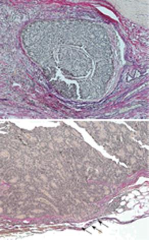
Figure 4 Histological features.
Nuclei of the endocrine cell carcinoma cells were irregular in size, and mitotis was frequently seen with extensive vein invasion partially penetrating the vein wall (arrows).
- Citation: Tanoue Y, Tanaka N, Suzuki Y, Hata S, Yokota A. A case report of endocrine cell carcinoma in the sigmoid colon with inferior mesenteric vein tumor embolism. World J Gastroenterol 2009; 15(2): 248-251
- URL: https://www.wjgnet.com/1007-9327/full/v15/i2/248.htm
- DOI: https://dx.doi.org/10.3748/wjg.15.248