Copyright
©2009 The WJG Press and Baishideng.
World J Gastroenterol. May 14, 2009; 15(18): 2265-2269
Published online May 14, 2009. doi: 10.3748/wjg.15.2265
Published online May 14, 2009. doi: 10.3748/wjg.15.2265
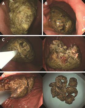
Figure 1 Endoscopic views of diospyrobezoar.
A: Initial endoscopic view. A huge dark brownish-colored diospyrobezoar was noted in the stomach (case 8). B: Endoscopic view of one day after 3 L of cola lavage. The size of bezoar was decreased. C: Endoscopic procedure.The remnant bezoar was captured and fragmented into four pieces by basket. D: Endoscopic procedure. The fragmented bezoar was crushed and retrieved by grasping force.
- Citation: Lee BJ, Park JJ, Chun HJ, Kim JH, Yeon JE, Jeen YT, Kim JS, Byun KS, Lee SW, Choi JH, Kim CD, Ryu HS, Bak YT. How good is cola for dissolution of gastric phytobezoars? World J Gastroenterol 2009; 15(18): 2265-2269
- URL: https://www.wjgnet.com/1007-9327/full/v15/i18/2265.htm
- DOI: https://dx.doi.org/10.3748/wjg.15.2265


