Copyright
©2009 The WJG Press and Baishideng.
World J Gastroenterol. May 14, 2009; 15(18): 2245-2251
Published online May 14, 2009. doi: 10.3748/wjg.15.2245
Published online May 14, 2009. doi: 10.3748/wjg.15.2245
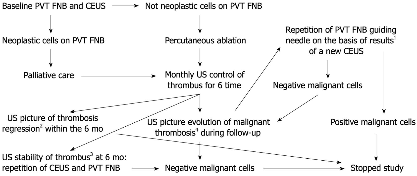
Figure 1 Study design.
PVT FNB: Portal vein thrombus fine needle biopsy; CEUS: 2nd generation Contrast-Enhanced US (CEUS) of thrombus; 1Guiding needle on portion of thrombus showing on CEUS precocious iso or hyperenhancement pattern; 2No increase in size and distribution with preservation of vessel wall or recanalization/shrinkage, or disappearance of a PVT within the 6 mo of follow-up were accepted as proof of a benign portal vein thrombus; 3No change in feature of thrombus and in the diameter of the segment of involved vein at 6 mo of follow-up; 4Increase in size with infiltration of perivascular parenchyma and interruption of vessel wall was US features indicative of malignant thrombosis.
-
Citation: Sorrentino P, D’Angelo S, Tarantino L, Ferbo U, Bracigliano A, Vecchione R. Contrast-enhanced sonography
versus biopsy for the differential diagnosis of thrombosis in hepatocellular carcinoma patients. World J Gastroenterol 2009; 15(18): 2245-2251 - URL: https://www.wjgnet.com/1007-9327/full/v15/i18/2245.htm
- DOI: https://dx.doi.org/10.3748/wjg.15.2245


