Copyright
©2009 The WJG Press and Baishideng.
World J Gastroenterol. Apr 14, 2009; 15(14): 1782-1785
Published online Apr 14, 2009. doi: 10.3748/wjg.15.1782
Published online Apr 14, 2009. doi: 10.3748/wjg.15.1782
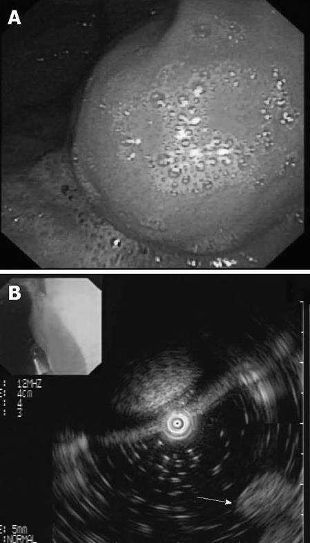
Figure 2 EUS was used to diagnose the gastric teratoma.
A: A spherical mass was noted on the inferior wall of the cardiac orifice, and the mass mucosa was normal; B: The heterogeneous mass was formed from the outer layer and the five-layer structure of the stomach in the mass was clearly detected (white arrow).
- Citation: Liu L, Zhuang W, Chen Z, Zhou Y, Huang XR. Primary gastric teratoma on the cardiac orifice in an adult. World J Gastroenterol 2009; 15(14): 1782-1785
- URL: https://www.wjgnet.com/1007-9327/full/v15/i14/1782.htm
- DOI: https://dx.doi.org/10.3748/wjg.15.1782


