Copyright
©2009 The WJG Press and Baishideng.
World J Gastroenterol. Apr 14, 2009; 15(14): 1719-1729
Published online Apr 14, 2009. doi: 10.3748/wjg.15.1719
Published online Apr 14, 2009. doi: 10.3748/wjg.15.1719
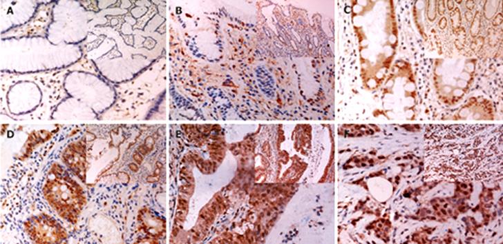
Figure 3 Immunohistochemical staining of AhR in gastric tissues.
A: CSG; B: CAG; C: IM; D: AH; E: i-GC; F: d-GC (Original magnification × 400 and × 200). Strong nuclear expression and weak cytoplasmic distribution of AhR were observed in epithelial cells and some stroma cells of both GC and pre-malignant tissues.
- Citation: Peng TL, Chen J, Mao W, Liu X, Tao Y, Chen LZ, Chen MH. Potential therapeutic significance of increased expression of aryl hydrocarbon receptor in human gastric cancer. World J Gastroenterol 2009; 15(14): 1719-1729
- URL: https://www.wjgnet.com/1007-9327/full/v15/i14/1719.htm
- DOI: https://dx.doi.org/10.3748/wjg.15.1719


