Copyright
©2009 The WJG Press and Baishideng.
World J Gastroenterol. Mar 21, 2009; 15(11): 1359-1366
Published online Mar 21, 2009. doi: 10.3748/wjg.15.1359
Published online Mar 21, 2009. doi: 10.3748/wjg.15.1359
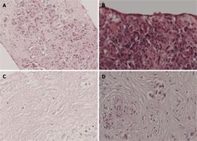
Figure 1 Histological staining of pancreatic tissue slices.
Slices derived from normal pancreas and pancreatic cancer were cultured for 3 d. A, B: Normal pancreas of good viability; C: Normal pancreas of poor viability showing massive tissue slice necrosis; D: Poorly differentiated adenocarcinoma of good viability. Hematoxylin/eosin staining. Original magnification of all tissues (100 ×).
-
Citation: Geer MAV, Kuhlmann KF, Bakker CT, Kate FJT, Elferink RPO, Bosma PJ.
Ex-vivo evaluation of gene therapy vectors in human pancreatic (cancer) tissue slices. World J Gastroenterol 2009; 15(11): 1359-1366 - URL: https://www.wjgnet.com/1007-9327/full/v15/i11/1359.htm
- DOI: https://dx.doi.org/10.3748/wjg.15.1359


