Copyright
©2008 The WJG Press and Baishideng.
World J Gastroenterol. Mar 7, 2008; 14(9): 1389-1393
Published online Mar 7, 2008. doi: 10.3748/wjg.14.1389
Published online Mar 7, 2008. doi: 10.3748/wjg.14.1389
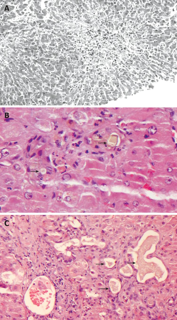
Figure 1 Histological findings in liver biopsy specimens.
A: Zone 3 (centrilobular) necrosis (HE, × 100); B: Canalicular cholestasis. Bile plugs in dilated canaliculi (black arrows). An apoptotic (acidophilic) body is present (white arrow) (HE, × 200); C: Ductular cholestasis and inflammation. Dilated bile ductules at the margin of an inflamed portal tract are filled with bile (black arrows) (HE, × 100).
- Citation: Koskinas J, Gomatos IP, Tiniakos DG, Memos N, Boutsikou M, Garatzioti A, Archimandritis A, Betrosian A. Liver histology in ICU patients dying from sepsis: A clinico-pathological study. World J Gastroenterol 2008; 14(9): 1389-1393
- URL: https://www.wjgnet.com/1007-9327/full/v14/i9/1389.htm
- DOI: https://dx.doi.org/10.3748/wjg.14.1389


