Copyright
©2008 The WJG Press and Baishideng.
World J Gastroenterol. Mar 7, 2008; 14(9): 1339-1345
Published online Mar 7, 2008. doi: 10.3748/wjg.14.1339
Published online Mar 7, 2008. doi: 10.3748/wjg.14.1339
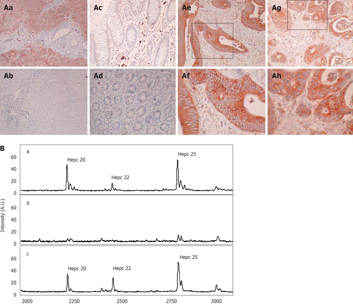
Figure 4 Cellular localization of hepcidin in colorectal cancer tissue.
A: To determine hepcidin cellular localization in colorectal tissue immunohistochemistry was performed with a hepcidin specific antibody (Abcam 31877). (Aa) Human Liver; (Ab) Human Liver, incubated with both hepcidin antibody and immunizing hepcidin peptide; (Ac) Normal colon; (Ad) Normal colon with crypts in cross section; (Ae) Colorectal cancer (x 20); (Af) Colorectal cancer (x 40); (Ag) colorectal cancer (x 20); (Ah) Colorectal cancer (x 40). Boxes denote area subsequently magnified; B: Immunocapture of urinary hepcidin. (Ba) A human urine sample containing hepcidin 20 (m/z 2193.6), 22 (m/z 2438.2) and 25 (m/z 2792.0) was subject to immunocapture with either (Bb) protein G sepharose or with (Bc) protein G-sepharose and a polyclonal anti hepcidin antibody. Immunocaptured proteins were then eluted from the protein G sepharose and analysed by MALDI-TOF MS.
- Citation: Ward DG, Roberts K, Brookes MJ, Joy H, Martin A, Ismail T, Spychal R, Iqbal T, Tselepis C. Increased hepcidin expression in colorectal carcinogenesis. World J Gastroenterol 2008; 14(9): 1339-1345
- URL: https://www.wjgnet.com/1007-9327/full/v14/i9/1339.htm
- DOI: https://dx.doi.org/10.3748/wjg.14.1339


