Copyright
©2008 The WJG Press and Baishideng.
World J Gastroenterol. Dec 14, 2008; 14(46): 7075-7085
Published online Dec 14, 2008. doi: 10.3748/wjg.14.7075
Published online Dec 14, 2008. doi: 10.3748/wjg.14.7075
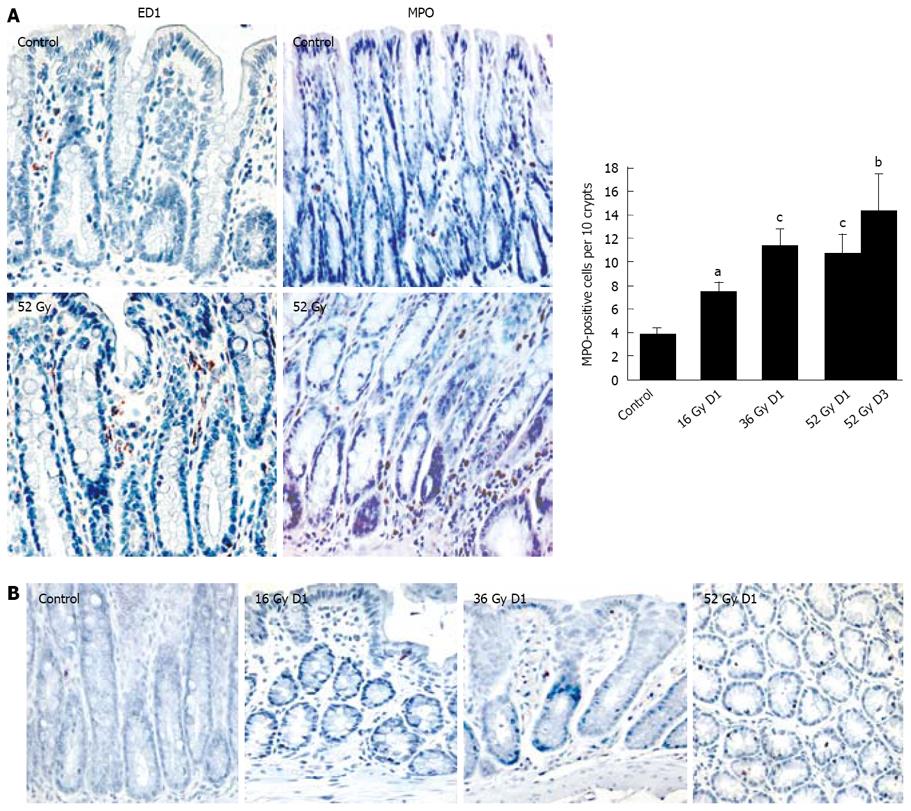
Figure 2 Effects of colorectal fractionated irradiation protocol on inflammatory cells and apoptotic cell presence.
Recruitment of ED-1-positive and MPO-positive cells (A) was assessed in distal colon mucosa in the normal distal colon (control) and three days after cumulative doses of 52 Gy. MPO-positive cells were correlated with the average number of neutrophils one day after cumulative doses of 16 Gy, 36 Gy, and one day and three days after 52 Gy. The presence of apoptotic cells was confirmed by the terminal deoxynucleotidyltransferase (TdT)-mediated dUTP-biotin nick-end labeling (TUNEL) staining at each timepoint of the fractionated protocol (B). Data are mean ± SE, aP < 0.01; bP < 0.005; cP < 0.001 vs control values. (× 20).
- Citation: Grémy O, Benderitter M, Linard C. Acute and persisting Th2-like immune response after fractionated colorectal γ-irradiation. World J Gastroenterol 2008; 14(46): 7075-7085
- URL: https://www.wjgnet.com/1007-9327/full/v14/i46/7075.htm
- DOI: https://dx.doi.org/10.3748/wjg.14.7075


