Copyright
©2008 The WJG Press and Baishideng.
World J Gastroenterol. Nov 21, 2008; 14(43): 6743-6747
Published online Nov 21, 2008. doi: 10.3748/wjg.14.6743
Published online Nov 21, 2008. doi: 10.3748/wjg.14.6743
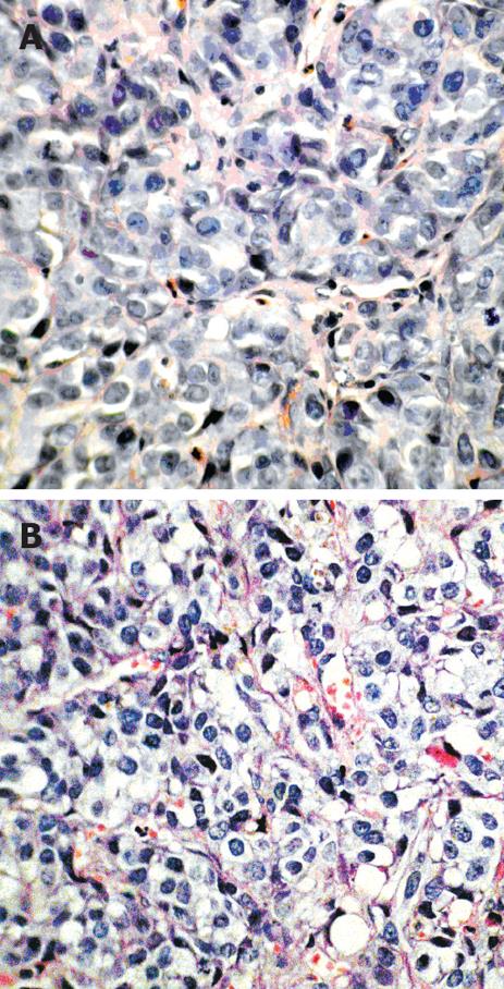
Figure 2 Light microscopic pathology of the targeted VX2 tumor rabbit liver tissues in sham group (A) and PHIFU + UCA group (B) (HE, x 400).
A: In sham group, tumor cells were large, irregularly arranged and with irregular morphology. The nuclei were large and deeply H&E stained, with great karyoplasmic ratio and increased mitosis. B: In PHIFU + UCA group, tumor cytoplasm in all 10 rabbits was lightly stained, with cytoplasmic vacuoles of various sizes, chromatin margination and karyopyknosis.
- Citation: Zhou CW, Li FQ, Qin Y, Liu CM, Zheng XL, Wang ZB. Non-thermal ablation of rabbit liver VX2 tumor by pulsed high intensity focused ultrasound with ultrasound contrast agent: Pathological characteristics. World J Gastroenterol 2008; 14(43): 6743-6747
- URL: https://www.wjgnet.com/1007-9327/full/v14/i43/6743.htm
- DOI: https://dx.doi.org/10.3748/wjg.14.6743


