Copyright
©2008 The WJG Press and Baishideng.
World J Gastroenterol. Nov 21, 2008; 14(43): 6711-6716
Published online Nov 21, 2008. doi: 10.3748/wjg.14.6711
Published online Nov 21, 2008. doi: 10.3748/wjg.14.6711
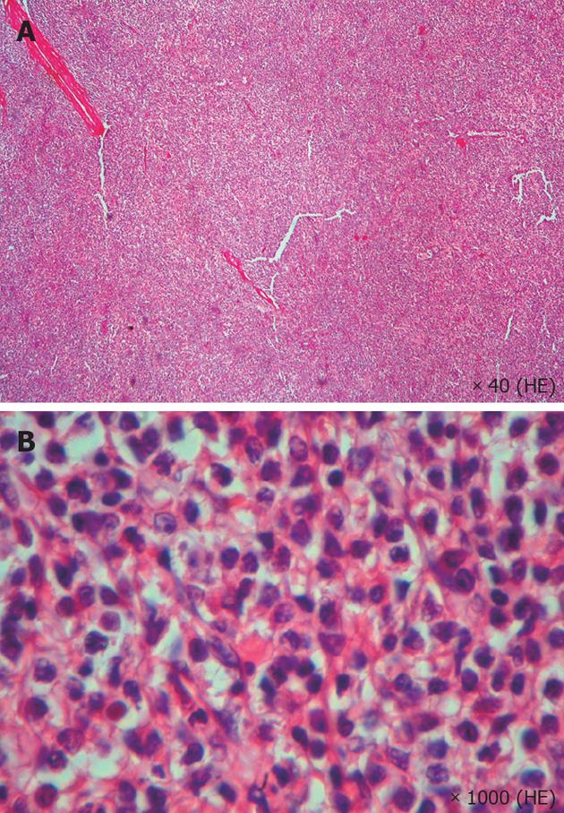
Figure 3 HE staining reveals splenic sinuses and cords of case VII (A, × 40) surrounded by hairy cells in the expanded red pulp (B, × 1000).
- Citation: Gedik E, Girgin S, Aldemir M, Keles C, Tuncer MC, Aktas A. Non-traumatic splenic rupture: Report of seven cases and review of the literature. World J Gastroenterol 2008; 14(43): 6711-6716
- URL: https://www.wjgnet.com/1007-9327/full/v14/i43/6711.htm
- DOI: https://dx.doi.org/10.3748/wjg.14.6711


