Copyright
©2008 The WJG Press and Baishideng.
World J Gastroenterol. Nov 14, 2008; 14(42): 6473-6480
Published online Nov 14, 2008. doi: 10.3748/wjg.14.6473
Published online Nov 14, 2008. doi: 10.3748/wjg.14.6473
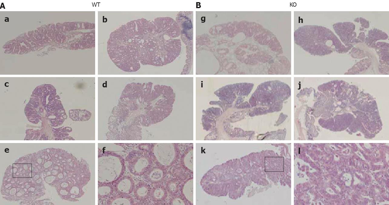
Figure 2 Histological analysis of colorectal tumors induced by AOM.
Representative HE-stained sections of colon tumors in WT mice (A): a: Adenoma; b, c, e: Carcinomas in situ; d: Adenocarcinoma; f: Boxed area in e is shown at a higher magnification. Representative HE-stained sections of colorectal tumors in KO mice (B). All tumors in KO mice showed features of adenocarcinoma: l: Boxed area in k is shown at a higher magnification. Original magnification: × 20 for b, c, d, h, i, j; × 40 for a, e, g, k; and × 200 for f and l.
- Citation: Nishihara T, Baba M, Matsuda M, Inoue M, Nishizawa Y, Fukuhara A, Araki H, Kihara S, Funahashi T, Tamura S, Hayashi N, Iishi H, Shimomura I. Adiponectin deficiency enhances colorectal carcinogenesis and liver tumor formation induced by azoxymethane in mice. World J Gastroenterol 2008; 14(42): 6473-6480
- URL: https://www.wjgnet.com/1007-9327/full/v14/i42/6473.htm
- DOI: https://dx.doi.org/10.3748/wjg.14.6473


