Copyright
©2008 The WJG Press and Baishideng.
World J Gastroenterol. Nov 7, 2008; 14(41): 6418-6420
Published online Nov 7, 2008. doi: 10.3748/wjg.14.6418
Published online Nov 7, 2008. doi: 10.3748/wjg.14.6418
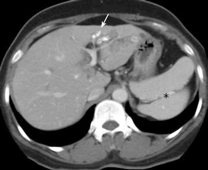
Figure 1 Abdominal computed tomography scan.
Polysplenia (asterisks) are present in the left upper quadrant and it shows the left lateral intrahepatic duct stones with atrophy (arrow).
- Citation: Seo HI, Jeon TY, Sim MS, Kim S. Polysplenia syndrome with preduodenal portal vein detected in adults. World J Gastroenterol 2008; 14(41): 6418-6420
- URL: https://www.wjgnet.com/1007-9327/full/v14/i41/6418.htm
- DOI: https://dx.doi.org/10.3748/wjg.14.6418


