Copyright
©2008 The WJG Press and Baishideng.
World J Gastroenterol. Jan 28, 2008; 14(4): 541-546
Published online Jan 28, 2008. doi: 10.3748/wjg.14.541
Published online Jan 28, 2008. doi: 10.3748/wjg.14.541
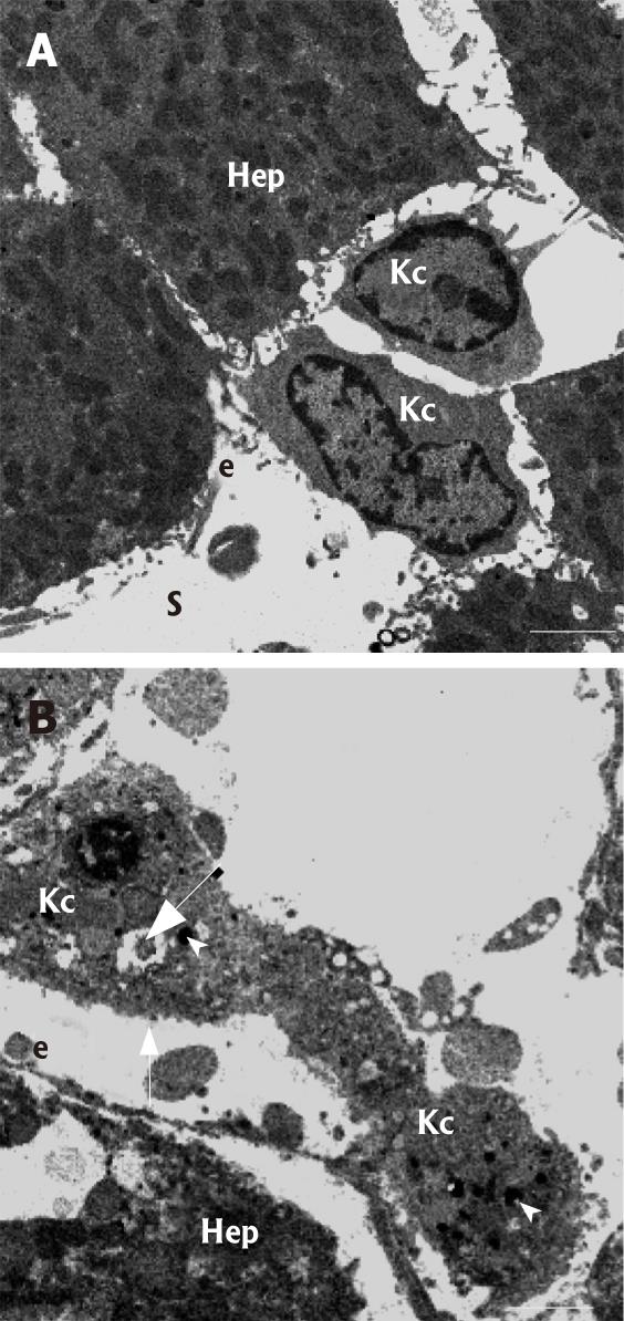
Figure 5 High-magnification transmission electron micro-graphs of Kupffer cells (Kc) in control (A) and kavain-treated livers (B).
A: Kupffer cells are located within the sinusoid (S) and adhere to the endothelial (e) lining by means of multiple cytoplasmic extensions. Note, hepatocytes (Hep); B: In kavain-treated livers, Kupffer cells (Kc) show an entire different ultrastructure when compared to control livers (see figure 4A for the difference): i.e., the cells look swollen, contain large amounts of electron dense phagocytosed material (arrowhead), have large vacuoles (large arrow), and have lost their ruffled and microvillous cell surface characteristics (small arrow). Scale bars, 5 micrometer.
- Citation: Fu S, Korkmaz E, Braet F, Ngo Q, Ramzan I. Influence of kavain on hepatic ultrastructure. World J Gastroenterol 2008; 14(4): 541-546
- URL: https://www.wjgnet.com/1007-9327/full/v14/i4/541.htm
- DOI: https://dx.doi.org/10.3748/wjg.14.541


