Copyright
©2008 The WJG Press and Baishideng.
World J Gastroenterol. Oct 21, 2008; 14(39): 6087-6092
Published online Oct 21, 2008. doi: 10.3748/wjg.14.6087
Published online Oct 21, 2008. doi: 10.3748/wjg.14.6087
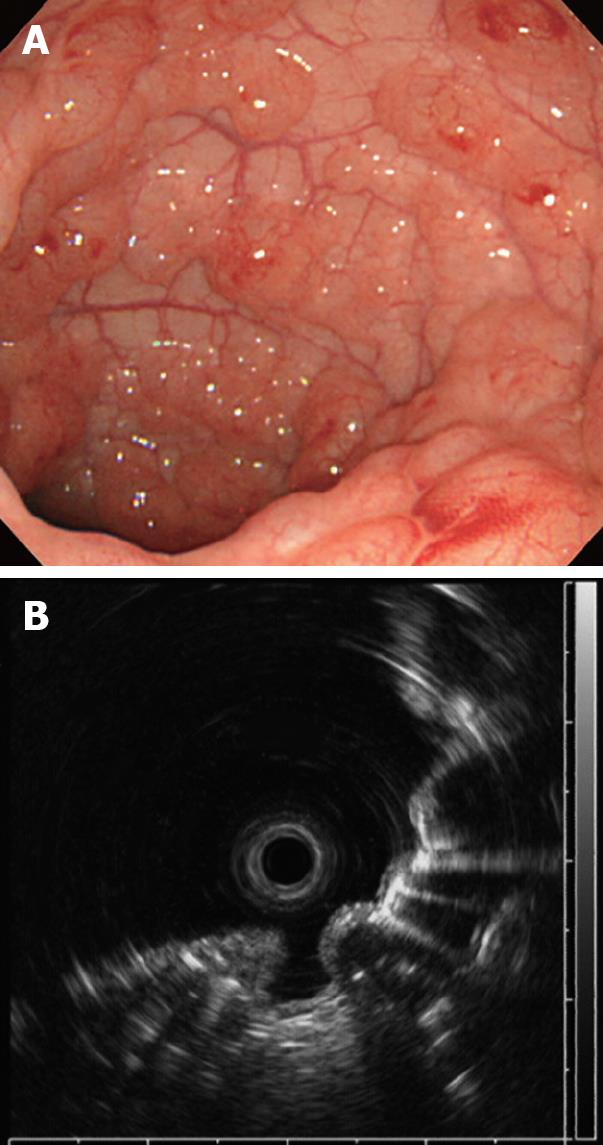
Figure 3 Colonoscopy on admission showing multiple round and smooth-surfaced elevated lesions like submucosal tumors in the sigmoid colon (A) and endoscopic ultrasonography (EUS) revealing hyperechoic lesions and acoustic shadows in the submucosal layer (B).
- Citation: Tsujimoto T, Shioyama E, Moriya K, Kawaratani H, Shirai Y, Toyohara M, Mitoro A, Yamao JI, Fujii H, Fukui H. Pneumatosis cystoides intestinalis following alpha-glucosidase inhibitor treatment: A case report and review of the literature. World J Gastroenterol 2008; 14(39): 6087-6092
- URL: https://www.wjgnet.com/1007-9327/full/v14/i39/6087.htm
- DOI: https://dx.doi.org/10.3748/wjg.14.6087


